

The development of high-throughput screening approaches for stem cell engineering
- PMID: 17702642
- PMCID: PMC3017753
- DOI: 10.1016/j.cbpa.2007.07.006
The development of high-throughput screening approaches for stem cell engineering
Abstract
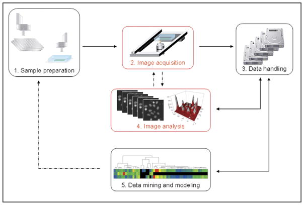
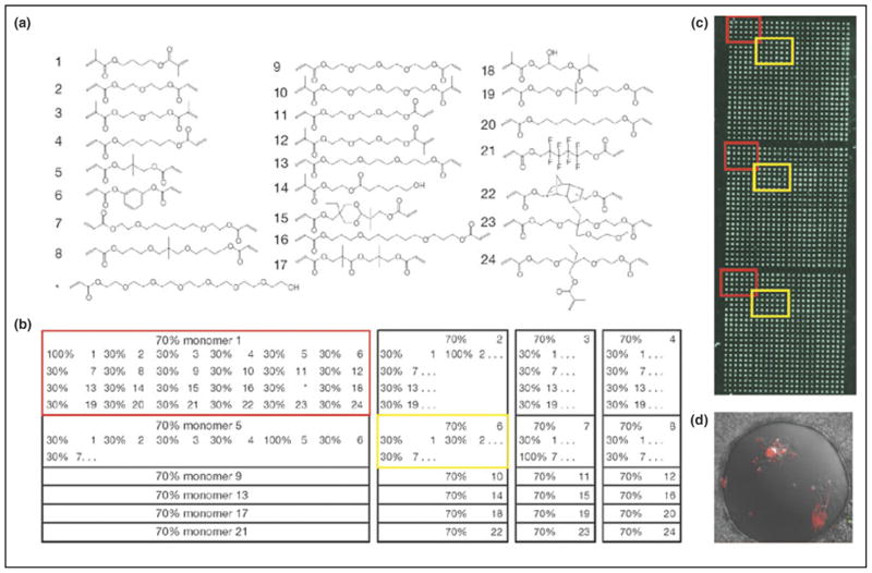
It has become increasingly clear that both soluble factors, such as growth factors, and insoluble factors, including the surfaces on which cells grow, can have controlling effects on stem cell behavior and differentiation. While much progress has been made in biomaterial design and application, the rational design of biomaterial cues to direct stem cell behavior and differentiation remains challenging. Recent advances in automated, high-throughput methods for synthesizing and screening combinatorial biomaterial libraries and cellular microenvironments promise to accelerate the discovery of factors that control stem cell behavior. Specific examples include miniaturized, automated, combinatorial material synthesis and extracellular matrix screening methods as well microarrayed methods for creating local microenvironments of soluble factors, such as small molecules, siRNA, and other signaling molecules.
Figures
Similar articles
-
High throughput optimization of stem cell microenvironments.Comb Chem High Throughput Screen. 2009 Jul;12(6):554-61. doi: 10.2174/138620709788681916. Comb Chem High Throughput Screen. 2009. PMID: 19601753 Free PMC article. Review.
-
Bioinspired materials for controlling stem cell fate.Acc Chem Res. 2010 Mar 16;43(3):419-28. doi: 10.1021/ar900226q. Acc Chem Res. 2010. PMID: 20043634 Free PMC article. Review.
-
Controlling stem cell fate with material design.Adv Mater. 2010 Jan 12;22(2):175-89. doi: 10.1002/adma.200901055. Adv Mater. 2010. PMID: 20217683
-
High throughput approaches for controlled stem cell differentiation.Acta Biomater. 2016 Apr 1;34:21-29. doi: 10.1016/j.actbio.2016.02.022. Epub 2016 Feb 13. Acta Biomater. 2016. PMID: 26884279 Review.
-
Combinatorial protein display for the cell-based screening of biomaterials that direct neural stem cell differentiation.Biomaterials. 2007 Feb;28(6):1048-60. doi: 10.1016/j.biomaterials.2006.10.004. Epub 2006 Nov 1. Biomaterials. 2007. PMID: 17081602
Cited by
-
Application of biomaterials to advance induced pluripotent stem cell research and therapy.EMBO J. 2015 Apr 15;34(8):987-1008. doi: 10.15252/embj.201490756. Epub 2015 Mar 12. EMBO J. 2015. PMID: 25766254 Free PMC article. Review.
-
Deeply dissecting stemness: making sense to non-coding RNAs in stem cells.Stem Cell Rev Rep. 2012 Mar;8(1):78-86. doi: 10.1007/s12015-011-9294-y. Stem Cell Rev Rep. 2012. PMID: 21706141 Review.
-
Multiplexed, high-throughput analysis of 3D microtissue suspensions.Integr Biol (Camb). 2010 Oct;2(10):517-27. doi: 10.1039/c0ib00054j. Epub 2010 Sep 1. Integr Biol (Camb). 2010. PMID: 20820630 Free PMC article.
-
Control of cultured human cells with femtosecond laser ablated patterns on steel and plastic surfaces.Biomed Microdevices. 2013 Apr;15(2):279-88. doi: 10.1007/s10544-012-9726-8. Biomed Microdevices. 2013. PMID: 23179464 Free PMC article.
-
Microarrayed Materials for Stem Cells.Mater Today (Kidlington). 2012 Oct 1;15(10):10.1016/S1369-7021(12)70196-7. doi: 10.1016/S1369-7021(12)70196-7. Mater Today (Kidlington). 2012. PMID: 24311967 Free PMC article.
References
-
- Polak J, Bishop A. Stem cells and tissue engineering: past, present, and future. Ann NY Acad Sci. 2006;1068:352–366. - PubMed
-
- Tutter A, Baltus G, Kadam S. Embryonic stem cells: a great hope for a new era of medicine. Curr Opin Drug Discov Dev. 2006;9:169–175. - PubMed
-
- Verfaillie CM, Pera MF, Lansdorp PM. Stem cells: hype and reality. Hematology. 2002;2002:369–391. - PubMed
-
- Odorico JS, Kaufman DS, Thomson JA. Multilineage differentiation from human embryonic stem cell lines. Stem Cells. 2001;19:193–204. - PubMed
-
- Thomson JA, Itskovitz-Eldor J, Shapiro SS, Waknitz MA, Swiergiel JJ, Marshall VS, Jones JM. Embryonic stem cell lines derived from human blastocysts. Science. 1998;282:1145–1147. - PubMed
Publication types
MeSH terms
Substances
Grants and funding
LinkOut - more resources
Full Text Sources
Other Literature Sources
Medical
