. 2007;35(16):5439-51.
doi: 10.1093/nar/gkm594. Epub 2007 Aug 15.

Protein/DNA arrays identify nitric oxide-regulated cis-element and trans-factor activities some of which govern neuroblastoma cell viability

Affiliations

Protein/DNA arrays identify nitric oxide-regulated cis-element and trans-factor activities some of which govern neuroblastoma cell viability

Saravanakumar Dhakshinamoorthy et al. Nucleic Acids Res. 2007.

Abstract

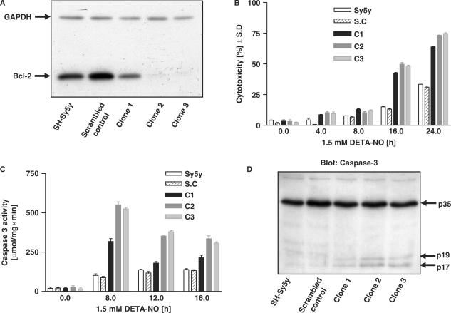
Toxic nitric oxide (NO) levels can regulate gene expression. Using a novel protein/DNA array, we show that toxic NO levels regulate the binding of trans-factors to various cis-elements in neuroblastoma cells, including CRE and those recognized by the transcription factors AP1, AP2, Brn-3a, EGR, E2F1 and SP1. Functionality of some of the cis-elements was confirmed by electro mobility shift and reporter assays. Interestingly, CREB, AP-1, Brn-3a, EGR and E2F1 can control mammalian cell viability. NO induced the anti-apoptotic Bcl-2 protein and its mRNA prior to the onset of death of 30-60% of the cells. Promoter analysis of the bcl-2 gene confirmed the involvement of a CRE in NO-dependent bcl-2 transcription. Neuroblastoma cells over-expressing bcl-2 became much more resistant to NO-induced apoptosis; conversely, Bcl-2 knockdown cells were rendered markedly more sensitive to NO. Together these results suggest that Bcl-2 counteracts NO-induced apoptosis in a fraction of the cell population. Thus, NO stimulates the binding of many trans-factors to their cognate cis-elements, some of which can regulate cell viability through transcriptional activation of target genes. Our results emphasize that a DNA/protein array approach can reveal novel, global transcription factor activities stimulated by cell death-regulating molecules.


Figures

Figure 1.
Figure 1.
NO induces apoptosis in SH-Sy5y Cells. NO donor-induced cell death was quantitated using Crystal Violet staining or by measuring caspase-3 activity. (A) SH-Sy5y cells were untreated or treated with SNP (2 mM) or DETA-NO (1.5 mM) or media alone (control) and stained using Crystal Violet. The results are represented as percentage cell death. (B) Apoptosis was measured by caspase-3 activation in SH-Sy5y cells treated with SNP or DETA-NO. In each of panels A and B, the values represent mean ± SE of three independent experiments.
Figure 2.
Figure 2.
NO induces the binding of trans-factors to an array of cis-elements. (A) SH-Sy5y cells were treated with SNP (2 mM) and the DNA/protein hybrid array (Panomics) was performed as described in Materials and Methods section. Detection of selected cis-elements is shown. (B and C) NO enhances the binding of nuclear factors to the various cis-elements. The cis-elements were end-labeled with γ-32P ATP. A total of 50 000 c.p.m. of the labeled cis-elements was incubated with 10 µg of nuclear extract from SH-Sy5y cells treated with the indicated NO donors for various times and analyzed in a 5% non-denaturing polyacrylamide gel. Shifted DNA–nuclear protein complexes are arrowed. (D) Competition assay. The wild-type (WT) cis-elements were end-labeled with γ-32P ATP. A total of 50 000 c.p.m. of labeled cis-elements were incubated with 10 µg of nuclear extract from SH-Sy5y cells treated with SNP either in the absence (control) or presence of 5×, 10× and 20× excess cold wild type (cold WT) or cold mutant (cold MT) oligonucleotide. The samples were analyzed in a 5% non-denaturing polyacrylamide gel. Shifted DNA–nuclear protein complexes are arrowed.
Figure 3.
Figure 3.
Effect of NO on transcriptional activation/repression mediated by the various cis-elements. Various cis-elements were cloned into the pGL3 vector with luciferase under the control of the SV40 promoter. SH-Sy5y cells were transfected with 0.5 µg of the various reporter plasmids along with 0.05 µg of the plasmid pRL-TK encoding Renilla luciferase. At 36 h post-transfection, the cells were treated with the NO donor SNP (2 mM) for the indicated times and analyzed for luciferase activity. The values represent mean ± SE of three independent transfection experiments.
Figure 4.
Figure 4.
Effect of NO on the expression of transcription factors. (A and B) SH-Sy5y cells were treated with the NO donor SNP (2 mM) for 4 and 8 h, and the transcription factor array was performed as described in the Materials and Methods section. The subsequent expression profiles of the selected transcription factors (representing mRNA levels) are shown.
Figure 5.
Figure 5.
Bcl-2 gene is a transcriptional target of NO. (A) Semi-quantitative RT-PCR analysis by agarose gel electrophoresis. SH-Sy5y cells were treated with the NO donors DETA-NO, SNP or SIN1 in the indicated concentrations for various times, and semi-quantitative RT-PCR analysis was performed using 2 μg of total RNA for the bcl-2, bid and bcl-xL genes. β-actin is shown as a loading control. (B) Western blot analysis. SH-Sy5y cells were treated with 1.5 mM DETA-NO or 2 mM SNP for various times. Twenty microgram of total protein were loaded on a 10% polyacrylamide gel. The membrane was probed with a Bcl-2 monoclonal antibody (lower panels). The signals on the western blots were scanned, normalized against β-actin and represented as optical density to show the fold induction for bcl-2 at each time point (upper panels).
Figure 6.
Figure 6.
cAMP response element (CRE) mediates transcriptional regulation of the bcl-2 gene by NO. (A) Schematic map of the human bcl-2 gene promoter constructs. The full-length human bcl-2 gene promoter harbors two well characterized regions, P1 and P2 (29). The LB 322 construct harbors the P1 and P2 regions upstream of the full-length promoter. LB 335 contains only the P2 region, whereas LB 124 contains only the P1 region. LB 334 contains a partial region of P1 harboring one CRE element and two SP1 elements. LB 595 is same as LB 334 with a mutation in the CRE site. LB 1263 and LB 1283 are same as LB 334 with LB 1263 harboring a mutation in the proximal SP1 site and LB 1283 harboring a mutation in the distal SP1 site, respectively. (B) Transcriptional regulation of the bcl-2 gene by NO is mediated by the P1 region only. SH-Sy5y cells were transfected with 0.5 µg of the various reporter plasmids and 0.05 µg of the plasmid pRL-TK encoding Renilla luciferase. At 36 h post-transfection, the cells were treated with DETA-NO (1.5 mM) for the indicated times and analyzed for luciferase activity. (C) Transcriptional regulation of bcl-2 gene by NO is mediated by a CRE element in the P1 region. SH-Sy5y cells were transfected with the various reporter plasmids, treated with DETA-NO (1.5 mM) and analyzed for luciferase activity as in B above. (D) Same as panel C, except that SNP (2 mM) was used instead of DETA-NO as NO donor. In each of panels B, C and D, the values represent mean ± SE of three independent transfection experiments. ▾1—distal Sp1 site, ▾2—CRE site, ▾3—proximal sp1 site, X—denotes mutation in the respective site.
Figure 7.
Figure 7.
Stable cells over-expressing bcl-2 are highly resistant to NO-induced apoptosis. (A) SH-Sy5y cells were transfected with the plasmid pcDNA-Bcl-2-myc or the pcDNA vector to generate stable cells. Left panels, similar levels of expression of the bcl-2-myc over-expressed proteins (single arrows) in the stable clones (Clone #1, Clone #2) were visualized by western blot analysis. Right panels, western blot comparing NO-induced caspase-3 cleavage in the wild-type cells (WT), vector control (Vector) and the pcDNA-Bcl-2-myc stable clones (Clone #1, Clone #2). (B) SH-Sy5y wild-type cells (WT), vector control (Vector) and bcl-2 stable clones (#1, #2) were treated with the NO donors SNP (2 mM) or DETA-NO (1.5 mM) for the indicated times, and NO-induced cell death was quantitated using the LDH release assay. (C) SH-Sy5y wild-type cells (WT), vector control (Vector) and bcl-2 stable clones (#1, #2) were treated with the NO donors SNP (2 mM) or DETA-NO (1.5 mM) for the indicated times, and apoptosis was assessed by measuring caspase-3 activity.
Figure 8.
Figure 8.
Stable knockdown of Bcl-2 sensitizes SH-Sy5y cells to NO-induced cell death. (A) Identification of stable Bcl-2 knockdown clones. Total proteins were subjected to electrophoresis in 0.1% SDS–14% polyacrylamide gels and western blot analysis for Bcl-2. Three individual clones (C1, C2 and C3) were analyzed. (B) SH-Sy5y parental cells, scrambled Bcl-2 oligonucleotide control cells (S.C) and three different clones of shBcl-2-expressing cells (C1–C3) were treated with 1.5 mM DETA-NO for the indicated times, and cytotoxicity was measured by LDH release. (C) As in B, except total proteins were harvested and caspase-3 activity was measured. (D) SH-Sy5y parental cells, vector control cells and three different clones of shBcl-2-expressing cells were treated with 1.5 mM DETA-NO for 16 h, and total proteins were subjected to electrophoresis as above followed by western blot analysis to detect procaspase-3 (p35) and cleaved caspase-3 fragments (p17, p19). Values in panels B and C are mean ± S.D determined from three experiments performed in triplicate.

References

    1. Michel T, Feron O. Nitric oxide synthases: which, where, how, and why? J. Clin. Invest. 1997;100:2146–2152. - PMC - PubMed
    1. Bogdan C. The multiplex function of nitric oxide in (auto)immunity. J. Exp. Med. 1998;187:1361–1365. - PMC - PubMed
    1. Hemish J, Nakaya N, Mittal V, Enikolopov G. Nitric oxide activates diverse signaling pathways to regulate gene expression. J. Biol. Chem. 2003;278:42321–42329. - PubMed
    1. Moncada S, Erusalimsky JD. Does nitric oxide modulate mitochondrial energy generation and apoptosis? Nat. Rev. Mol. Cell. Biol. 2002;3:214–220. - PubMed
    1. Lucas KA, Pitari GM, Kazerounian S, Ruiz-Stewart I, Park J, Schulz S, Chepenik KP, Waldman SA. Guanylyl cyclases and signaling by cyclic GMP. Pharmacol. Rev. 2000;52:375–414. - PubMed

Publication types