. 2007 Dec 1;110(12):3996-4004.
doi: 10.1182/blood-2007-02-074450. Epub 2007 Aug 16.

Establishment of transplantable porcine tumor cell lines derived from MHC-inbred miniature swine

Affiliations

Establishment of transplantable porcine tumor cell lines derived from MHC-inbred miniature swine

Patricia S Cho et al. Blood. .

Abstract

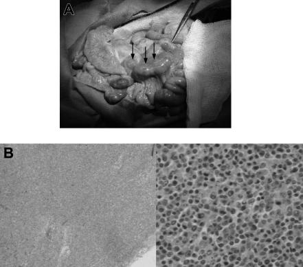
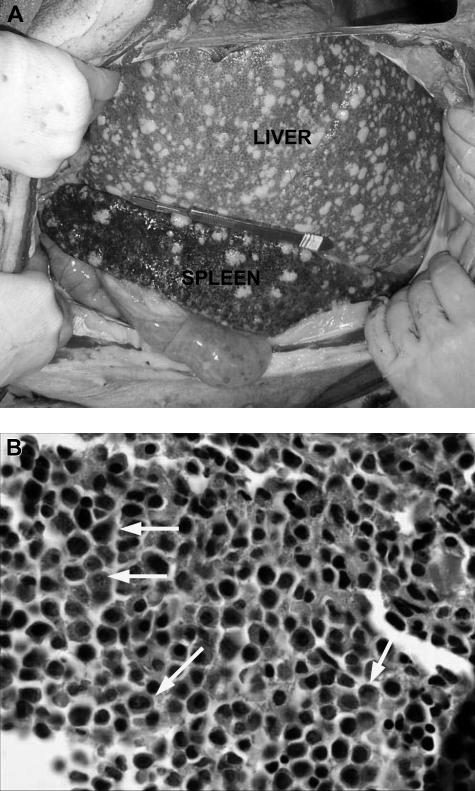
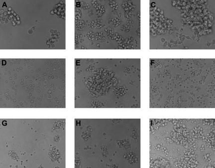
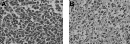
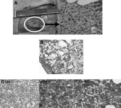
The lack of transplantable tumors has limited assessment of graft-versus-tumor effects following hematopoietic cell transplantation in clinically relevant large-animal models. We describe the derivation and characterization of porcine tumor cell lines with initial efforts of tumor transplantation using immunocompromised mice and highly inbred sublines of Massachusetts General Hospital major histocompatibility complex (MHC)-inbred miniature swine. Autopsies were performed routinely on swine that died unexpectedly or had suspicion of malignancy based on clinical symptoms or peripheral blood analysis. Tissue samples were obtained for pathology, phenotyped by flow cytometry, and placed in culture. Based on growth, lines were selected for passage into nonobese diabetic/severe combined immunodeficient (NOD/SCID) mice and miniature swine. Porcine tumor recipients were preconditioned with total body irradiation from 0 to 500 cGy or with a 30-day course of oral cyclosporine. We identified 19 cases of hematologic tumors. Nine distinct tumor cell lines were established from 8 of these cases, including 3 derived from highly inbred sublines. In vivo tumor growth and serial transfer were observed in immunocompromised mice for one tumor cell line and in miniature swine for 1 of 2 tumor cell lines expanded for this purpose. These results suggest the possibility of developing a transplantable tumor model in this large-animal system.


Figures

Figure 1
Figure 1
Gross pathologic and histologic findings of lymphoma (PTLD) in miniature swine. (A) Animals that developed lymphomas or PTLD typically had pronounced lymphadenopathy, as represented by animal 17018. Image was acquired using a Kodak camera (Eastman Kodak, Rochester, NY) model Easyshare Z740 with a 45.5-mm to 55-mm lens adapter. No further image processing was done. (B) Lymph node tissue harvested from these animals demonstrated destruction of normal architecture and predominance of abnormal cells as represented by animal 13271. Slides were viewed with an Olympus BX51 compound microscope (Olympus America, Melville, NY) of sections stained with hematoxylin and eosin (H&E; Hematoxylin Gill's Formulation no. 2, Fisher Diagnostics, Fair Lawn, NJ; Eosin-Y, Richard-Allan Scientific, Kalamazoo, MI) using a lens at 40× (left) and 400× (right). Images were acquired using an Olympus digital microscope camera (Olympus America) model Q-Color 3, and were processed with Adobe Photoshop CS version 8 software (Adobe Systems, San Jose, CA).
Figure 2
Figure 2
Gross pathologic and histologic findings of leukemia in miniature swine. (A) The most consistent findings of animals with leukemias were enlarged liver and spleen, which on palpation were firm and pale in color with visible lesions, as shown by animal 15549. Image was acquired using a Kodak camera (Eastman Kodak) model Easyshare Z740 with a 45.5-mm to 55-mm lens adapter. No further image processing was done. (B) Bone marrow from these animals was predominantly populated with abnormal cells, as represented by tissue from animal 14736. Slide was viewed with an Olympus BX51 compound microscope (Olympus America) of sections stained with H&E (Hematoxylin Gill's Formulation no. 2, Fisher Diagnostics; Eosin-Y, Richard-Allan Scientific) using a lens at 400×. Image was acquired using an Olympus digital microscope camera (Olympus America) model Q-Color 3, and was processed with Adobe Photoshop CS version 8 software (Adobe Systems).
Figure 3
Figure 3
In vitro growth of tumor cell lines. The 9 tumor cell lines display various patterns of growth and sizes in vitro. Most preferentially grow in clusters, although one line, 14736, grows as a single-cell suspension. (A) MML-12933; (B) LCL-13271; (C) ML-13381; (D) CML-14736; (E) CML-15433; (F) LCL-15446; (G) LCL-17016L; (H) LCL-17016P; (I) LCL-17018. Slides were viewed with a Nikon Eclipse TE2000-U microscope (Nikon Instruments, Melville, NY) using Nikon Plan Fluor lenses at 40× in a cell culture media. Images were acquired using a NIkon Eclipse TE2000-U camera (Nikon Instruments) and were processed with Adobe Photoshop Elements version 3.0 software (Adobe Systems).
Figure 4
Figure 4
Histologic findings after in vivo transfer of LCL-13271 tumor cells in NOD/SCID mice. LCL-13271 injected intraperitoneally into NOD/SCID mice with tumor growth at 2 months in primary and secondary recipients. Abdominal tumor mass from NOD/SCID primary (A) and secondary (B) recipients of LCL-13271. Histologic findings were similar in morphology compared with the primary tumor (Huang et al). Slides were viewed with an Olympus BX40 microscope (Olympus America) of sections stained with H&E medium (Hematoxylin Gill's Formulation no. 2, Fisher Diagnostics; Eosin-Y, Richard-Allan Scientific) using a lens at 400×. Images were acquired using a Hitachi charge-coupled device color camera (Hitachi Kokusai Electric America, Woodbury, NY) model HV-C20 3-CCD, and were processed with ACDSee version 4.0 software (ACD Systems International, Victoria, BC).
Figure 5
Figure 5
Histologic findings after in vivo transfer of CML-14736 tumor cells into miniature swine. CML-14736 grew after in vivo transfer to histocompatible miniature swine pretreated with TBI. Tumor growth was found at the subcutaneous injection sites (A) and in the lungs after intravenous administration (B). Immunohistochemistry of the subcutaneous injection site tissue negative staining for CD3, but positive staining for CD16 and CD172a, which was consistent with the surface phenotype of the primary tumor and cultured cells (C). Slides were veiwed with a Nikon Eclipse E800 microscope (Nikon Instruments) of sections stained with H&E (Hematoxylin Gill's Formulation no. 2, Fisher Diagnostics; Eosin-Y, Richard-Allan Scientific) using Nikon Plan Fluor lenses at 400× (A right; C left, middle, right) and 200× (B). Images were acquired using Nikon HRD060-NIK 0.6X optical coupler diagnostic instruments (Nikon Instruments) connected to a computer with SPOT-diagnostic instruments and was processed with SPOT Advanced or Basic version 3.5.6 software for Windows (Diagnostic Instruments, Sterling Heights, MI).

References

    1. Adelman CA, Petrini JH, Attwooll CL. Modeling disease in the mouse: lessons from DNA damage response and cell cycle control genes. J Cell Biochem. 2006;97:459–473. - PubMed
    1. Braithwaite AW, Royds JA, Jackson P. The p53 story: layers of complexity. Carcinogenesis. 2005;26:1161–1169. - PubMed
    1. Decker S, Sausville EA. Preclinical modeling of combination treatments: fantasy or requirement? Ann N Y Acad Sci. 2005;1059:61–69. - PubMed
    1. Egorov IK. Mouse models of efficient and inefficient anti-tumor immunity, with emphasis on minimal residual disease and tumor escape. Cancer Immunol Immunother. 2006;55:1–22. - PMC - PubMed
    1. Gutmann DH, Hunter-Schaedle K, Shannon KM. Harnessing preclinical mouse models to inform human clinical cancer trials. J Clin Invest. 2006;116:847–852. - PMC - PubMed

Publication types

MeSH terms