. 2007 Sep 14;317(5844):1544-8.
doi: 10.1126/science.1142819. Epub 2007 Aug 16.

Crystal structure of an ancient protein: evolution by conformational epistasis

Affiliations

Crystal structure of an ancient protein: evolution by conformational epistasis

Eric A Ortlund et al. Science. .

Abstract

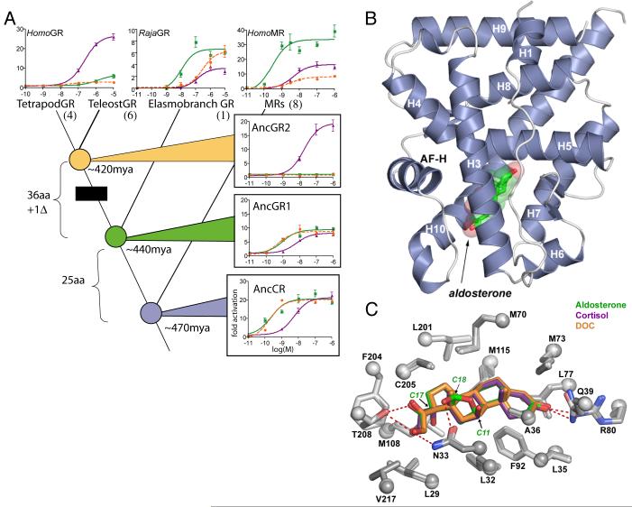
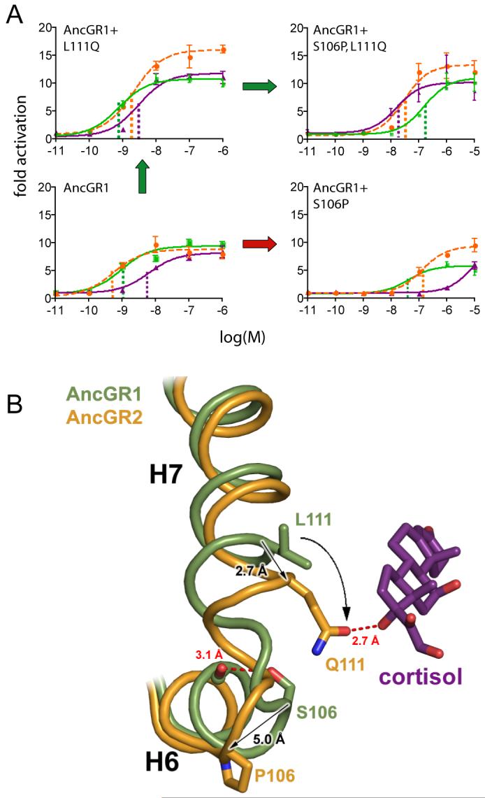
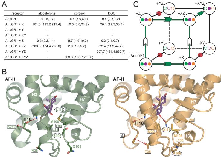
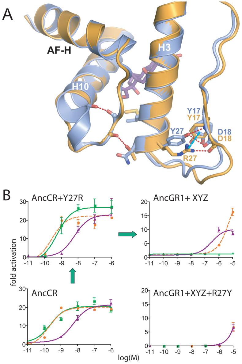
The structural mechanisms by which proteins have evolved new functions are known only indirectly. We report x-ray crystal structures of a resurrected ancestral protein-the approximately 450 million-year-old precursor of vertebrate glucocorticoid (GR) and mineralocorticoid (MR) receptors. Using structural, phylogenetic, and functional analysis, we identify the specific set of historical mutations that recapitulate the evolution of GR's hormone specificity from an MR-like ancestor. These substitutions repositioned crucial residues to create new receptor-ligand and intraprotein contacts. Strong epistatic interactions occur because one substitution changes the conformational position of another site. "Permissive" mutations-substitutions of no immediate consequence, which stabilize specific elements of the protein and allow it to tolerate subsequent function-switching changes-played a major role in determining GR's evolutionary trajectory.


Figures

Fig. 1
Fig. 1
A) Functional evolution of corticosteroid receptors. Dose-response curves show transcription of a luciferase reporter gene by extant and resurrected ancestral receptors with varying doses (in log-M) of aldosterone (green), deoxycorticosterone (DOC, orange), and cortisol (purple). Black box indicates evolution of cortisol specificity. The number of sequence changes on each branch is shown (aa, replacement; Δ deletion). Bars, SEM of three replicates. Node dates from the fossil record (19, 20). For complete phylogeny and sequences, see Figs S10 and Table S5. B) Crystal structure of the AncCR ligand-binding domain (LBD) with bound aldosterone (green, with red oxygens). Helices are labeled; AF-H, activation-function helix. C) AncCR’s ligand-binding pocket. Side chains (<4.2 Å from bound ligand) are superimposed from crystal structures of AncCR with aldosterone (green), DOC (orange), and cortisol (purple). Oxygen and nitrogen atoms are red and blue; dashed lines, hydrogen bonds. Arrows show C11, C17, and C18 positions, which differ among the hormones.
Fig. 2
Fig. 2
Mechanism for switching AncGR1’s ligand preference from aldosterone to cortisol. A) Effect of substitutions S106P and L111Q on the resurrected AncGR1’s response to hormones. Dashed lines indicate sensitivity to aldosterone (green), cortisol (purple), and DOC (orange) as the EC50 (concentration at which half-maximal reporter activation is achieved). Green arrow shows probable pathway through a functional intermediate; red arrow, intermediate with radically reduced sensitivity to all hormones. B) Structural change conferring new ligand specificity. Backbones of helices 6 and 7 from AncGR1 (green) and AncGR2 (yellow) in complex with cortisol are superimposed. Substitution S106P induces a kink in the interhelical loop of AncGR2, repositioning sites 106 and 111 (arrows). In this background, L111Q forms a new hydrogen bond with cortisol’s unique C17-hydroxyl (dotted red line).
Fig. 3
Fig. 3
Permissive substitutions in the evolution of receptor specificity. A) Effects of various combinations of historical substitutions on AncGR1’s transcriptional activity and hormone-response in a reporter gene assay. Group Y (L29M, F98I, and S212Δ) abolishes receptor activity unless groups X (S106P, L111Q) and Z (N26T and Q105L) are present; the XYZ combination yields complete cortisol-specificity. Table shows hormone sensitivity as the EC50 (in nM). Parentheses, 95% confidence interval. Dash, no activation. B) Structural prediction of permissive substitutions. Models of AncGR1 (green) and AncGR2 (yellow) are shown with cortisol. Group X and Y substitutions (circles and rectangles) yield new interactions with the C17-hydroxyl of cortisol (purple) but destabilize receptor regions required for activation. Group Z (underlined) impart additional stability to the destabilized regions. C) Restricted evolutionary paths through sequence space. Nodes of the cube represent states for residue sets X, Y, and Z. Edges represent pathways from the ancestral sequence (AncGR1) to the cortisol-specific combination (+XYZ). Filled circles at vertices show sensitivity to aldosterone (green), DOC (orange), and cortisol (purple); empty circles, no activation. Red octagons, paths through non-functional intermediates; arrows, paths through functional intermediates with no change (white) or switched ligand preference (green).
Fig. 4
Fig. 4
Structural identification of an ancient permissive substitution. A) Comparison of the structures of AncCR (blue) and AncGR2 (yellow). Y27R generates a novel cation-π interaction in AncGR2 (dotted cyan line), replacing the weaker ancestral hydrogen bond (dotted red) and imparting additional stability to helix 3. B) Y27R is permissive for the substitutions that confer GR function. Reporter gene activation by AncGR1+XYZ (upper right) is abolished when Y27R is reversed (lower right). Left, Y27R has negligible effect in the AncCR background (or in AncGR1,Fig. S9). Green, orange, and purple lines show aldosterone, DOC, and cortisol response. Green arrows, likely pathway through functional intermediates.

References

    1. Golding GB, Dean AM. Mol Biol Evol. 1998;15:355. - PubMed
    1. Stern DL. Evolution Int J Org Evolution. 2000;54:1079. - PubMed
    1. Perutz MF. Mol Biol Evol. 1983;1:1. - PubMed
    1. Glasner ME, Gerlt JA, Babbitt PC. Curr Opin Chem Biol. 2006;10:492. - PubMed
    1. Kinch LN, Grishin NV. Curr Opin Struct Biol. 2002;12:400. - PubMed

Publication types

MeSH terms