

Comparison of computed tomographic angiography versus rubidium-82 positron emission tomography for the detection of patients with anatomical coronary artery disease
- PMID: 17703259
- PMCID: PMC2651386
- DOI: 10.1016/s0828-282x(07)70831-0
Comparison of computed tomographic angiography versus rubidium-82 positron emission tomography for the detection of patients with anatomical coronary artery disease
Abstract
Background: The present study compared computed tomographic coronary angiography (CTA) and positron emission tomography (PET) for the detection of significant anatomical coronary artery stenosis as defined by conventional invasive coronary angiography (CICA).
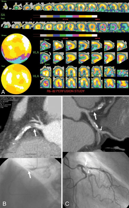
Methods: The study protocol was approved by the local ethics board, and informed consent was obtained from all patients. Of the 26 patients (mean age 57+/-9 years, 18 men) who prospectively underwent CTA and rubidium-82 PET before CICA, 24 patients had a history of chest pain. Images were interpreted by expert readers and assessed for the presence of anatomically significant coronary stenosis (50% luminal diameter stenosis or greater) or myocardial perfusion defects. Diagnostic test characteristics were analyzed using patient-based, territory-based, vessel-based and segment-based analyses.
Results: In the 24 patients referred for chest pain, CTA had similar sensitivity to PET, but was more specific (sensitivity 95% [95% CI 72% to 100%] versus 95% [95% CI 72% to 100%], respectively; specificity 100% [95% CI 46% to 100%] versus 60% [95% CI 17% to 93%], respectively) in the detection of patients with anatomical coronary artery stenosis of 50% or greater. On a per-segment basis of all 26 patients, CTA had a sensitivity, specificity, positive predictive value and negative predictive value of 72%, 99%, 91% and 95%, respectively, in all coronary segments.
Conclusions: Coronary CTA has a similar sensitivity and specificity to rubidium-82 PET for the identification of patients with significant anatomical coronary artery disease.
CONTEXTE :: La présente étude visait à comparer la coronarographie par tomodensitométrie (CTDM) avec la tomographie par émission de positrons (TEP) en vue de la détection de sténoses anatomiques coronariennes importantes, confirmées par la coronarographie classique effractive (CCE).
MÉTHODE :: Le protocole de l’étude a reçu l’approbation du comité local d’éthique, et tous les sujets ont signé un consentement éclairé. Sur 26 patients (âge moyen : 57±9 ans; hommes : 18) qui ont subi, de façon prospective, la CTDM et la TEP au rubidium 82 avant la CCE, 24 avaient des antécédents de douleur thoracique. Les images ont été interprétées par des spécialistes, qui les ont examinées à la recherche de sténoses anatomiques coronariennes importantes, c’est-à-dire égales ou supérieures à 50 % de la lumière des vaisseaux, ou d’images lacunaires d’irrigation. Il y a eu analyse des caractéristiques des examens de diagnostic en fonction des patients, des territoires, des vaisseaux et des segments.
RÉSULTATS :: Chez les 24 patients dirigés en radiologie pour des douleurs thoraciques, la CTDM avait une sensibilité comparable à celle de la TEP mais une spécificité plus grande que celle-ci (sensibilité : 95 % [IC : 72 % à 100 %] contre 95 % [IC : 72 % à 100 %], respectivement; spécificité : 100 % [IC : 46 % à 100 %] contre 60 % [IC : 17 % à 93 %], respectivement) en ce qui concerne la détection des sténoses anatomiques coronariennes, égales ou supérieures à 50 %. Quant à l’analyse par segment chez les 26 sujets, la CTDM avait une sensibilité, une spécificité, une valeur prédictive positive et une valeur prédictive négative de 72 %, 99 %, 91 % et 95 % respectivement, et ce, dans tous les segments coronariens.
CONCLUSION :: La CTDM a une sensibilité et une spécificité comparables à celles de la TEP au rubidium 82 pour la détection des sténoses anatomiques coronariennes importantes.
Figures
Similar articles
-
Detection of significant coronary artery disease by noninvasive anatomical and functional imaging.Circ Cardiovasc Imaging. 2015 Mar;8(3):e002179. doi: 10.1161/CIRCIMAGING.114.002179. Circ Cardiovasc Imaging. 2015. PMID: 25711274 Clinical Trial.
-
Diagnostic accuracy of rubidium-82 myocardial perfusion imaging with hybrid positron emission tomography/computed tomography in the detection of coronary artery disease.J Am Coll Cardiol. 2007 Mar 13;49(10):1052-8. doi: 10.1016/j.jacc.2006.12.015. Epub 2007 Feb 26. J Am Coll Cardiol. 2007. PMID: 17349884
-
Diagnostic Performance of Coronary CT Angiography and Myocardial Perfusion Imaging in Kidney Transplantation Candidates.JACC Cardiovasc Imaging. 2015 May;8(5):553-562. doi: 10.1016/j.jcmg.2014.12.028. Epub 2015 Apr 10. JACC Cardiovasc Imaging. 2015. PMID: 25869350
-
Non-invasive imaging in coronary artery disease including anatomical and functional evaluation of ischaemia and viability assessment.Br J Radiol. 2011 Dec;84 Spec No 3(Spec Iss 3):S280-95. doi: 10.1259/bjr/50903757. Br J Radiol. 2011. PMID: 22723535 Free PMC article. Review.
-
Developments in coronary CT angiography.Curr Cardiol Rep. 2008 Feb;10(1):51-9. doi: 10.1007/s11886-008-0011-7. Curr Cardiol Rep. 2008. PMID: 18417002 Review.
Cited by
-
Diagnosing coronary artery disease with hybrid PET/CT: it takes two to tango.J Nucl Cardiol. 2013 Oct;20(5):874-90. doi: 10.1007/s12350-013-9753-8. J Nucl Cardiol. 2013. PMID: 23842709 Review.
-
Influence of coronary artery disease prevalence on predictive values of coronary CT angiography: a meta-regression analysis.Eur Radiol. 2011 Sep;21(9):1904-13. doi: 10.1007/s00330-011-2142-2. Epub 2011 May 20. Eur Radiol. 2011. PMID: 21597986 Review.
-
Myocardial flow reserve (MFR) with positron emission tomography (PET)/computed tomography (CT): clinical impact in diagnosis and prognosis.Cardiovasc Diagn Ther. 2017 Apr;7(2):206-218. doi: 10.21037/cdt.2017.04.10. Cardiovasc Diagn Ther. 2017. PMID: 28540215 Free PMC article. Review.
-
Combined evaluation of regional coronary artery calcium and myocardial perfusion by 82Rb PET/CT in the identification of obstructive coronary artery disease.Eur J Nucl Med Mol Imaging. 2018 Apr;45(4):521-529. doi: 10.1007/s00259-018-3935-1. Epub 2018 Jan 25. Eur J Nucl Med Mol Imaging. 2018. PMID: 29372272
-
Responsible use of computed tomography in the evaluation of coronary artery disease and chest pain.Mayo Clin Proc. 2010 Apr;85(4):358-64. doi: 10.4065/mcp.2009.0652. Mayo Clin Proc. 2010. PMID: 20360294 Free PMC article. Review.
References
-
- Schuijf JD, Bax JJ, Shaw LJ, et al. Meta-analysis of comparative diagnostic performance of magnetic resonance imaging and multislice computed tomography for noninvasive coronary angiography. Am Heart J. 2006;151:404–11. - PubMed
-
- Hoffmann U, Moselewski F, Cury RC, et al. Predictive value of 16-slice multidetector spiral computed tomography to detect significant obstructive coronary artery disease in patients at high risk for coronary artery disease: Patient-versus segment-based analysis. Circulation. 2004;110:2638–43. - PubMed
-
- Hoffmann MH, Shi H, Schmitz BL, et al. Noninvasive coronary angiography with multislice computed tomography JAMA 20052932471–8.(Erratum in 2005;294:1208). - PubMed
-
- Mollet NR, Cademartiri F, Nieman K, et al. Multislice spiral computed tomography coronary angiography in patients with stable angina pectoris. J Am Coll Cardiol. 2004;43:2265–70. - PubMed
-
- Kuettner A, Trabold T, Schroeder S, et al. Noninvasive detection of coronary lesions using 16-detector multislice spiral computed tomography technology: Initial clinical results. J Am Coll Cardiol. 2004;44:1230–7. - PubMed
