

Structural change and nucleotide dissociation of Myosin motor domain: dual go model simulation
- PMID: 17704146
- PMCID: PMC2084243
- DOI: 10.1529/biophysj.106.103796
Structural change and nucleotide dissociation of Myosin motor domain: dual go model simulation
Abstract
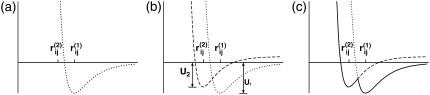
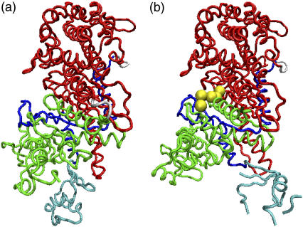
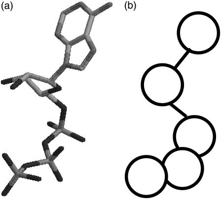
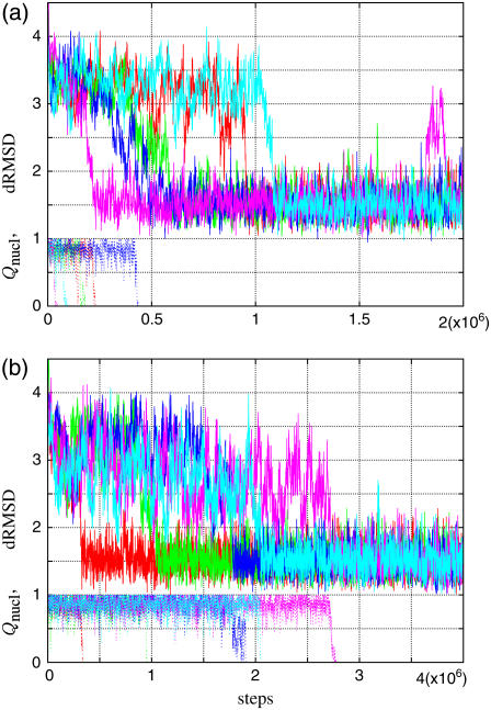
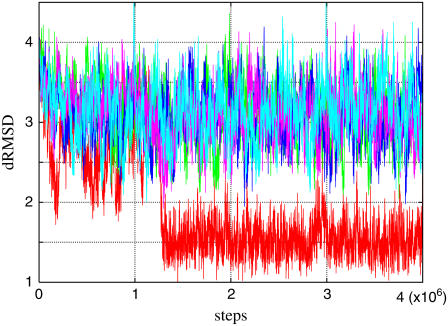
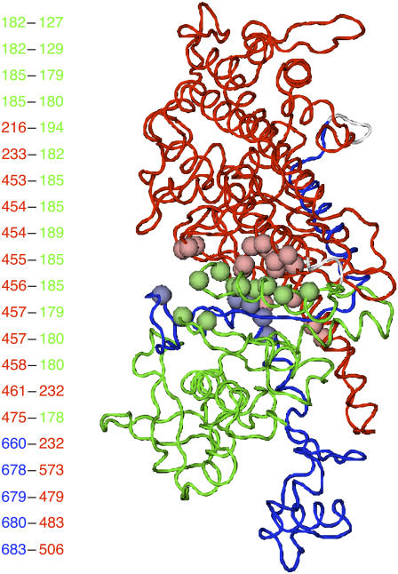
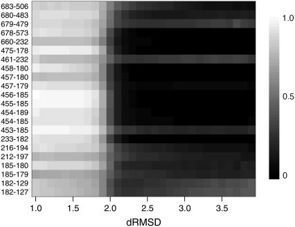
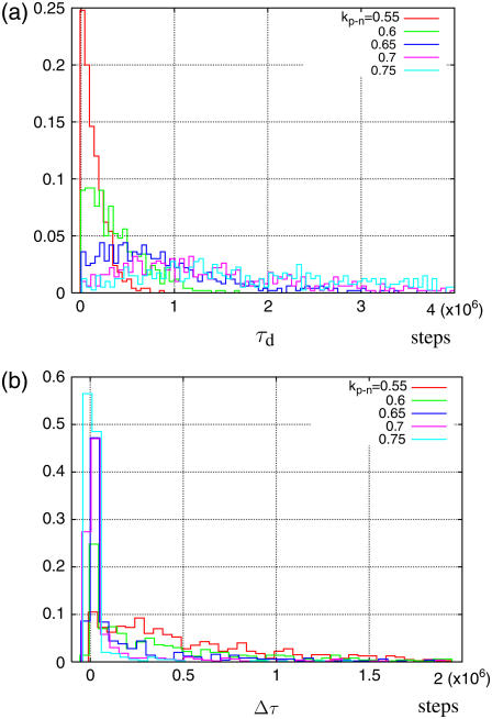
We investigated the structural relaxation of myosin motor domain from the pre-power stroke state to the near-rigor state using molecular dynamics simulation of a coarse-grained protein model. To describe the spontaneous structural change, we propose a dual Gō-model-a variant of the Gō-like model that has two reference structures. The nucleotide dissociation process is also studied by introducing a coarse-grained nucleotide in the simulation. We found that the myosin structural relaxation toward the near-rigor conformation cannot be completed before the nucleotide dissociation. Moreover, the relaxation and the dissociation occurred cooperatively when the nucleotide was tightly bound to the myosin head. The result suggested that the primary role of the nucleotide is to suppress the structural relaxation.
Figures
Similar articles
-
Mutation in the SH1 helix reduces the activation energy of the ATP-induced conformational transition of myosin.Biochem Biophys Res Commun. 2007 May 25;357(1):325-9. doi: 10.1016/j.bbrc.2007.03.155. Epub 2007 Apr 2. Biochem Biophys Res Commun. 2007. PMID: 17416346
-
Thiol reactivity as a sensor of rotation of the converter in myosin.Biochem Biophys Res Commun. 2008 Apr 25;369(1):115-23. doi: 10.1016/j.bbrc.2007.11.148. Epub 2007 Dec 7. Biochem Biophys Res Commun. 2008. PMID: 18068118
-
Structure and dynamics of the force-generating domain of myosin probed by multifrequency electron paramagnetic resonance.Biophys J. 2008 Jul;95(1):247-56. doi: 10.1529/biophysj.107.124305. Epub 2008 Mar 13. Biophys J. 2008. PMID: 18339764 Free PMC article.
-
Calcium and cargoes as regulators of myosin 5a activity.Biochem Biophys Res Commun. 2008 Apr 25;369(1):176-81. doi: 10.1016/j.bbrc.2007.11.109. Epub 2007 Dec 3. Biochem Biophys Res Commun. 2008. PMID: 18060865 Review.
-
Cooperativity of myosin molecules through strain-dependent chemistry.Philos Trans R Soc Lond B Biol Sci. 2000 Apr 29;355(1396):529-38. doi: 10.1098/rstb.2000.0594. Philos Trans R Soc Lond B Biol Sci. 2000. PMID: 10836506 Free PMC article. Review.
Cited by
-
Insights from coarse-grained Gō models for protein folding and dynamics.Int J Mol Sci. 2009 Mar;10(3):889-905. doi: 10.3390/ijms10030889. Epub 2009 Mar 2. Int J Mol Sci. 2009. PMID: 19399227 Free PMC article. Review.
-
Building a macro-mixing dual-basin Gō model using the Multistate Bennett Acceptance Ratio.Biophys Physicobiol. 2019 Nov 29;16:310-321. doi: 10.2142/biophysico.16.0_310. eCollection 2019. Biophys Physicobiol. 2019. PMID: 31984186 Free PMC article.
-
Entropic mechanism of large fluctuation in allosteric transition.Proc Natl Acad Sci U S A. 2010 Apr 27;107(17):7775-80. doi: 10.1073/pnas.0912978107. Epub 2010 Apr 12. Proc Natl Acad Sci U S A. 2010. PMID: 20385843 Free PMC article.
-
Coevolution of function and the folding landscape: correlation with density of native contacts.Biophys J. 2008 Nov 1;95(9):L57-9. doi: 10.1529/biophysj.108.143388. Epub 2008 Aug 15. Biophys J. 2008. PMID: 18708465 Free PMC article.
-
Effects of ATP and actin-filament binding on the dynamics of the myosin II S1 domain.Biophys J. 2013 Oct 1;105(7):1624-34. doi: 10.1016/j.bpj.2013.08.023. Biophys J. 2013. PMID: 24094403 Free PMC article.
References
-
- Geeves, M. A., and K. C. Holmes. 1999. Structural mechanism of muscle contraction. Annu. Rev. Biochem. 68:687–728. - PubMed
-
- Spudich, J. A. 2001. The myosin swinging cross-bridge model. Nat. Rev. Mol. Cell Biol. 2:387–392. - PubMed
-
- Cruz, E. M. D. L., and E. M. Ostap. 2004. Relating biochemistry and function in the myosin superfamily. Curr. Opin. Cell Biol. 16:61–67. - PubMed
Publication types
MeSH terms
Substances
LinkOut - more resources
Full Text Sources
