. 2007 Dec 1;93(11):3860-71.
doi: 10.1529/biophysj.107.112060. Epub 2007 Aug 17.

Coarse-grained free energy functions for studying protein conformational changes: a double-well network model

Affiliations

Coarse-grained free energy functions for studying protein conformational changes: a double-well network model

Jhih-Wei Chu et al. Biophys J. .

Abstract

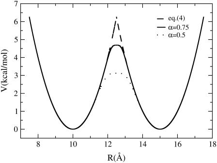
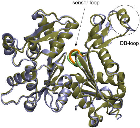
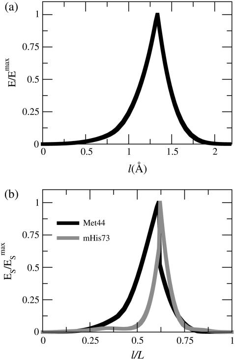
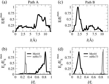
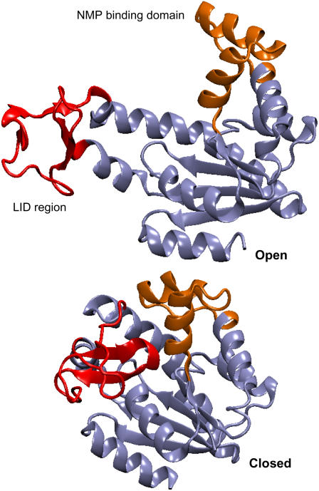
In this work, a double-well network model (DWNM) is presented for generating a coarse-grained free energy function that can be used to study the transition between reference conformational states of a protein molecule. Compared to earlier work that uses a single, multidimensional double-well potential to connect two conformational states, the DWNM uses a set of interconnected double-well potentials for this purpose. The DWNM free energy function has multiple intermediate states and saddle points, and is hence a "rough" free energy landscape. In this implementation of the DWNM, the free energy function is reduced to an elastic-network model representation near the two reference states. The effects of free energy function roughness on the reaction pathways of protein conformational change is demonstrated by applying the DWNM to the conformational changes of two protein systems: the coil-to-helix transition of the DB-loop in G-actin and the open-to-closed transition of adenylate kinase. In both systems, the rough free energy function of the DWNM leads to the identification of distinct minimum free energy paths connecting two conformational states. These results indicate that while the elastic-network model captures the low-frequency vibrational motions of a protein, the roughness in the free energy function introduced by the DWNM can be used to characterize the transition mechanism between protein conformations.


Figures

FIGURE 1
FIGURE 1
The combined energy profile in kcal/mol/Å of interpolating two one-dimensional harmonic potentials by using the PNM and DWNM approaches. The equilibrium positions for the two potentials are located at 10 and 15 Å, respectively. Both potentials have a force constant of 1 kcal/mol/Å. The solid curve was obtained by PNM (Eq. 4), the dashed curve was obtained by DWNM (Eq. 6), with α = 0.75, and the dotted curve was obtained by DWNM (Eq. 6), with α = 0.5.
FIGURE 2
FIGURE 2
The x-ray structures of ATP-bound (G-ATP, blue, PDB code No. 1NWK) and ADP-bound (G-ADP, green, PDB code No. 1J6Z) G-actin. The sensor-loop and the DB-loop are highlighted. The sensor-loop is colored orange for G-ATP and light green for G-ADP. The DB-loop assumes a coiled structure in G-ATP while folding into an α-helix in G-ADP.
FIGURE 3
FIGURE 3
Properties along the minimum free energy path (MFEP) connecting G-ATP and G-ADP structures by applying the PNM. (a) The normalized free energy as a function of path length. The energies are normalized by the maximum value along the path. (b) The normalized strain energy of Met44 in the DB-loop and mHis73 in the sensor-loop as a function of the normalized path length.
FIGURE 4
FIGURE 4
Properties along the MFEP connecting G-ATP and G-ADP structures by applying the DWNM. Path A was obtained by a linearly interpolated initial structure and Path B was obtained by using the PNM MFEP as the initial structure for MEP optimization. (a) The normalized energy as a function of the path length of Path A. The energies are normalized by the maximum value of energy of Path B. (b) The normalized strain energy of Met44 in the DB-loop and mHis73 in the sensor-loop as a function of the normalized path length of Path A. (c) The normalized energy as a function of the path length of Path B. The energies are normalized by the maximum value of energy of Path B. (d) The normalized strain energy of Met44 in the DB-loop and mHis73 in the sensor-loop as a function of the normalized path length of Path B.
FIGURE 5
FIGURE 5
The x-ray structures of the open-state conformation (PDB code No. 4AKE) and the closed-state conformation (PDB code No. 1AKE) of adenylate kinase. The LID region is colored in red and the NMP-binding region is colored in orange.
FIGURE 6
FIGURE 6
Properties along the MFEP connecting the open- and closed-state conformations of AKE by using the PNM. (a) The normalized energy as a function of path length. The energies are normalized by the maximum value of the path. (b) The values of (dod)/(dodc) between residues 127 (LID region) and 194 (CORE domain) and between residues 55 (NMP-binding domain) and 169 (CORE domain) as a function of the normalized path length.
FIGURE 7
FIGURE 7
Properties along the MFEP connecting the open- and closed-state conformations of AKE by using the DWNM. Path A was obtained by a linearly interpolated initial structure and Path B was obtained by using the MFEP obtained by the PNM. (a) The normalized energy as a function of the path length of Path A. The energies are normalized by the maximum value of energy of Path A. (b) The values of (dod)/(dodc) between residues 127 (LID region) and 194 (CORE domain) and between residues 55 (NMP-binding domain) and 169 (CORE domain) as a function of the normalized path length of Path A. (c) The normalized energy as a function of the path length of Path B. The energies are normalized by the maximum value of energy of Path A. (d) The values of (dod)/(dodc) between residues 127 (LID region) and 194 (CORE domain) and between residues 55 (NMP-binding domain) and 169 (CORE domain) as a function of the normalized path length of Path B.

Similar articles

Cited by

References

    1. Gao, Z. G., and K. A. Jacobson. 2006. Allosterism in membrane receptors. Drug Discov. Today. 11:191–202. - PMC - PubMed
    1. Karplus, M., and J. Kuriyan. 2005. Molecular dynamics and protein function. Proc. Natl. Acad. Sci. USA. 102:6679–6685. - PMC - PubMed
    1. Lodish, H., M. P. Scott, P. Matsudaira, J. Darnell, L. Zipursky, C. A. Kaiser, A. Berk, and M. Krieger. 2003. Molecular Cell Biology. W. H. Freeman, New York.
    1. Ackers, G. K. 1998. Deciphering the molecular code of hemoglobin allostery. Adv. Protein Chem. 51:185–253. - PubMed
    1. Crivici, A., and M. Ikura. 1995. Molecular and structural basis of target recognition by calmodulin. Annu. Rev. Biophys. Biomol. Struct. 24:85–116. - PubMed

Publication types

LinkOut - more resources