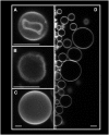
Giant unilamellar vesicles electroformed from native membranes and organic lipid mixtures under physiological conditions
- PMID: 17704162
- PMCID: PMC2072068
- DOI: 10.1529/biophysj.107.116228
Giant unilamellar vesicles electroformed from native membranes and organic lipid mixtures under physiological conditions
Abstract
In recent years, giant unilamellar vesicles (GUVs) have become objects of intense scrutiny by chemists, biologists, and physicists who are interested in the many aspects of biological membranes. In particular, this "cell size" model system allows direct visualization of particular membrane-related phenomena at the level of single vesicles using fluorescence microscopy-related techniques. However, this model system lacks two relevant features with respect to biological membranes: 1), the conventional preparation of GUVs currently requires very low salt concentration, thus precluding experimentation under physiological conditions, and 2), the model system lacks membrane compositional asymmetry. Here we show for first time that GUVs can be prepared using a new protocol based on the electroformation method either from native membranes or organic lipid mixtures at physiological ionic strength. Additionally, for the GUVs composed of native membranes, we show that membrane proteins and glycosphingolipids preserve their natural orientation after electroformation. We anticipate our result to be important to revisit a vast variety of findings performed with GUVs under low- or no-salt conditions. These studies, which include results on artificial cell assembly, membrane mechanical properties, lipid domain formation, partition of membrane proteins into lipid domains, DNA-lipid interactions, and activity of interfacial enzymes, are likely to be affected by the amount of salt present in the solution.
Figures


Similar articles
-
Electroformation of giant unilamellar vesicles from native membranes and organic lipid mixtures for the study of lipid domains under physiological ionic-strength conditions.Methods Mol Biol. 2010;606:105-14. doi: 10.1007/978-1-60761-447-0_9. Methods Mol Biol. 2010. PMID: 20013393
-
Electroformation of giant unilamellar vesicles from erythrocyte membranes under low-salt conditions.Anal Biochem. 2013 Apr 15;435(2):174-80. doi: 10.1016/j.ab.2013.01.001. Epub 2013 Jan 17. Anal Biochem. 2013. PMID: 23333270
-
Giant unilamellar vesicle electroformation from lipid mixtures to native membranes under physiological conditions.Methods Enzymol. 2009;465:161-76. doi: 10.1016/S0076-6879(09)65009-6. Methods Enzymol. 2009. PMID: 19913167
-
Giant unilamellar vesicles - a perfect tool to visualize phase separation and lipid rafts in model systems.Acta Biochim Pol. 2009;56(1):33-9. Epub 2009 Mar 17. Acta Biochim Pol. 2009. PMID: 19287805 Review.
-
Targeting membrane proteins to liquid-ordered phases: molecular self-organization explored by fluorescence correlation spectroscopy.Chem Phys Lipids. 2006 Jun;141(1-2):158-68. doi: 10.1016/j.chemphyslip.2006.02.026. Epub 2006 Apr 4. Chem Phys Lipids. 2006. PMID: 16696961 Review.
Cited by
-
Enzymatic determination of diglyceride using an iridium nano-particle based single use, disposable biosensor.Sensors (Basel). 2010;10(6):5758-73. doi: 10.3390/s100605758. Epub 2010 Jun 8. Sensors (Basel). 2010. PMID: 22219685 Free PMC article.
-
Preparation of size tunable giant vesicles from cross-linked dextran(ethylene glycol) hydrogels.Chem Commun (Camb). 2014 Feb 25;50(16):1953-5. doi: 10.1039/c3cc49144g. Chem Commun (Camb). 2014. PMID: 24407820 Free PMC article.
-
Biophysical approaches for exploring lipopeptide-lipid interactions.Biochimie. 2020 Mar;170:173-202. doi: 10.1016/j.biochi.2020.01.009. Epub 2020 Jan 21. Biochimie. 2020. PMID: 31978418 Free PMC article. Review.
-
Membrane organization and regulation of cellular cholesterol homeostasis.J Membr Biol. 2010 Apr;234(3):183-94. doi: 10.1007/s00232-010-9245-6. Epub 2010 Mar 25. J Membr Biol. 2010. PMID: 20336284 Free PMC article.
-
Quantitative analysis of the lamellarity of giant liposomes prepared by the inverted emulsion method.Biophys J. 2014 Jul 15;107(2):346-354. doi: 10.1016/j.bpj.2014.05.039. Biophys J. 2014. PMID: 25028876 Free PMC article.
References
-
- Singer, S. J., and G. L. Nicolson. 1972. The fluid mosaic model of the structure of cell membranes. Science. 175:720–731. - PubMed
-
- Edidin, M. 2003. The state of lipid rafts: from model membranes to cells. Annu. Rev. Biophys. Biomol. Struct. 32:257–283. - PubMed
-
- Simons, K., and W. L. Vaz. 2004. Model systems, lipid rafts, and cell membranes. Annu. Rev. Biophys. Biomol. Struct. 33:269–295. - PubMed
-
- Mukherjee, S., and F. R. Maxfield. 2004. Membrane domains. Annu. Rev. Cell Dev. Biol. 20:839–866. - PubMed
-
- Bagatolli, L. A. 2006. To see or not to see: lateral organization of biological membranes and fluorescence microscopy. Biochim. Biophys. Acta. 1758:1541–1556. - PubMed
Publication types
MeSH terms
Substances
LinkOut - more resources
Full Text Sources

