. 2007 Dec 1;93(11):4041-52.
doi: 10.1529/biophysj.107.109298. Epub 2007 Aug 17.

Simultaneous tether extraction from endothelial cells and leukocytes: observation, mechanics, and significance

Affiliations

Simultaneous tether extraction from endothelial cells and leukocytes: observation, mechanics, and significance

Gaurav Girdhar et al. Biophys J. .

Abstract

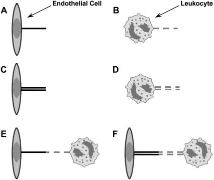
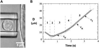
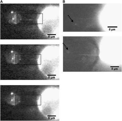
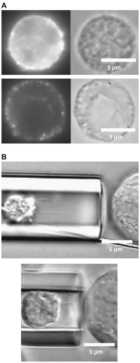
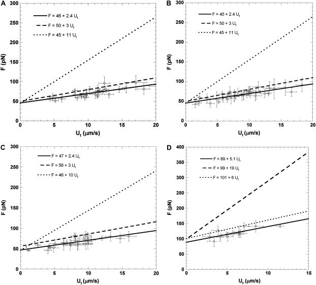
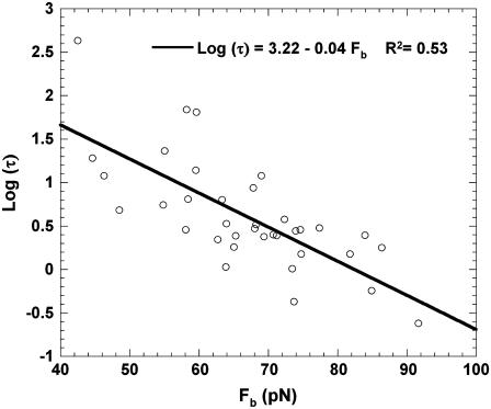
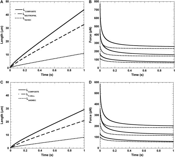
It has been hypothesized, from earlier studies on single-tether extraction from individual leukocytes and human umbilical vein endothelial cells, that during rolling of leukocytes on the endothelium, simultaneous extraction of membrane nanotubes (tethers) occurs, resulting in enhancement of the force decrease on the adhesive bond. In this study, using the micropipette aspiration technique and fluorescence microscopy, we show that tethers are indeed extracted simultaneously when an endothelial cell and a leukocyte are separated after brief contact and adhesion, and the endothelial cell contributes much more to the composite tether length. In addition, the constitutive relationship for simultaneous tether extraction is determined with neutrophils and T-lymphocytes as force transducers, and cytokine-stimulated human umbilical vein and dermal microvascular endothelial cells as substrates, respectively. This relationship is consistent with that derived theoretically from the constitutive equations for single-tether extraction from either cell alone. Moreover, we show that simultaneous tether extraction was likely terminated by receptor-ligand bond dissociation. With a biomechanical model of leukocyte rolling, we predict the force history of the adhesive receptor-ligand bond and show that it is remarkably similar for different leukocyte-endothelial cell pairs. Simultaneous tether extraction therefore represents a generic mechanism for stabilizing leukocyte rolling on the endothelium.


Figures

FIGURE 1
FIGURE 1
Six possible scenarios of tether extraction from endothelial cells and leukocytes in vitro or in vivo. (A and B) Single-tether extraction from endothelial cells (A) or leukocytes (B) alone. (C and D) Double-tether extraction from endothelial cells (C) or leukocytes (D) alone. (E and F) Single- (E) or double-tether (F) extraction from leukocytes and endothelial cells simultaneously.
FIGURE 2
FIGURE 2
The microscopic view of simultaneous tether extraction from leukocytes and attached ECs using MAT. (A) The experimental setup for simultaneous tether extraction from a surface-attached endothelial cell (TNF-α-treated HUVEC) and a passive leukocyte (neutrophil) as the force transducer. (B) The tracked displacement (D) of a leukocyte during a typical double-tether extraction event: (1) Approach toward the EC; (2) Contact with the EC; (3) Double tether being extracted; (4) Single tether being extracted; (5) Free motion of the leukocyte. The three fitted solid lines clearly show the distinct velocities during phases 3–5.
FIGURE 3
FIGURE 3
(A) Simultaneous tether extraction from a neutrophil (left) and a HUVEC (right), where both cells are labeled with DiO. The three-frame observation for a typical simultaneous tether event (upper, elongation; middle and lower, tether retraction after bond rupture) is shown. The rectangular areas in each frame indicate the region of interest for simultaneous and individual tethers. (B) Simultaneous tether extraction from a HUVEC and a neutrophil. The HUVEC, at right, is labeled with FM1-43 and the neutrophil, at left, is unlabeled. The dotted line (arrowhead) represents the approximate position of the neutrophil. (Note the larger contribution of the EC to the composite tether length.)
FIGURE 4
FIGURE 4
(A) Disruption of actin filaments in the neutrophil after treatment with 100 μM cytochalasin D. Both control (upper) and cytochalasin-treated (lower) neutrophils are stained with Phalloidin Alexa Fluor to observe the actin filaments. (B) Simultaneous tether extraction from a HUVEC, at right, and a neutrophil treated with 100 μM cytochalasin D, at left. Note the enhanced contribution of the thicker neutrophil tether to the composite tether length and the junction adhesive bond more or less in the middle.
FIGURE 5
FIGURE 5
Simultaneous single-tether extraction from (A) passive neutrophils and TNF-α (10 ng/ml, 4 h) treated surface-attached HUVECs in Ca2+/Mg2+ medium, (B) passive neutrophils and TNF-α (10 ng/ml, 4 h) treated surface-attached HUVECs in Mg2+/EGTA/anti-E-selectin medium, (C) passive T-lymphocytes and surface-attached HDMECs-n treated with IL-1β (10 ng/ml, 6 h) in Ca2+/Mg2+ medium, and (D) simultaneous double-tether extraction from passive neutrophils and TNF-α (10 ng/ml, 4 h) treated surface-attached HUVECs in Mg2+/EGTA/anti-E-selectin medium. Each point represents an average of 5–20 tethers. The threshold force (intercept) and the effective viscosity (slope/2π) for simultaneous tether extraction can be estimated from the linear regression. In A and B, the dashed line represents single-tether extraction from attached and TNF-α-stimulated HUVECs and the dotted line represents single-tether extraction from passive neutrophils. In C, the dashed line represents single-tether extraction from attached IL-1β-stimulated HDMECs-n and the dotted line represents single-tether extraction from CD4+ T-lymphocytes. In D, the dashed line represents double-tether extraction from passive neutrophils and the dotted line represents double-tether extraction from attached TNF-α-stimulated HUVECs.
FIGURE 6
FIGURE 6
The bond lifetime (τ) as a function of pulling force (Fb) for the LFA-1-ICAM-1 interaction. Simultaneous single-tether extraction was accomplished from passive neutrophils treated with Mg2+/EGTA and surface-attached HUVECs stimulated with TNF-α (10 ng/ml, 4 h). Each point is an average of multiple adhesion events. The error bars for each point have been removed for clarity.
FIGURE 7
FIGURE 7
Calculated tether lengths and force histories for simultaneous tether extraction from endothelial cells and leukocytes. (A) The calculated growth of the simultaneous single HUVEC-neutrophil tether, and the individual contributions from the neutrophil and HUVEC to the total tether length (solid line) are plotted for a shear rate of 270 s−1. The dashed and dotted lines represent the contributions from the neutrophil and HUVEC, respectively. An initial microvillus length of 0.35 μm (L0) was used in the computation. Note that L2 = L0 at time 0. (B) The calculated decrease in the bond force over time during simultaneous extraction of three single neutrophil-HUVEC tethers is plotted for shear rates of 100, 270, and 450 s−1 (dotted lines, bottom to top). The solid line represents the calculated force decrease for the case of tether extraction from the neutrophil alone and is plotted here for comparison with the simultaneous tether. (C) The calculated growth of the single simultaneous HDMEC-n-T-cell tether and the individual contributions from the T-cell and HDMEC-n to the total tether length (solid line) are plotted for a shear rate of 270 s−1. The dashed and dotted lines represent contributions from the T-cell and HDMEC-n, respectively. An initial microvillus length of 0.35 μm (L0) was used in the computation. Note that L2 = L0 at time 0. (D) The calculated decrease in the bond force over time during simultaneous extraction of three single T-cell-HDMEC-n tethers is plotted for shear rates of 100, 270, and 450 s−1 (dotted lines, bottom to top). The solid line represents the calculated force decrease for the case of tether extraction from the T-cell alone and is plotted here for comparison with the simultaneous tether.

Similar articles

Cited by

References

    1. Park, E. Y., M. J. Smith, E. S. Stropp, K. R. Snapp, J. A. DiVietro, W. F. Walker, D. W. Schmidtke, S. L. Diamond, and M. B. Lawrence. 2002. Comparison of PSGL-1 microbead and neutrophil rolling: microvillus elongation stabilizes P-selectin bond clusters. Biophys. J. 82:1835–1847. - PMC - PubMed
    1. Ramachandran, V., M. Williams, T. Yago, D. W. Schmidtke, and R. P. McEver. 2004. Dynamic alterations of membrane tethers stabilize leukocyte rolling on P-selectin. Proc. Natl. Acad. Sci. USA. 101:13519–13524. - PMC - PubMed
    1. Yago, T., A. Leppanen, H. Qiu, W. D. Marcus, M. U. Nollert, C. Zhu, R. D. Cummings, and R. P. McEver. 2002. Distinct molecular and cellular contributions to stabilizing selectin-mediated rolling under flow. J. Cell Biol. 158:787–799. - PMC - PubMed
    1. Evans, E., V. Heinrich, A. Leung, and K. Kinoshita. 2005. Nano- to microscale dynamics of p-selectin detachment from leukocyte interfaces. I. Membrane separation from the cytoskeleton. Biophys. J. 88:2288–2298. - PMC - PubMed
    1. Shao, J.-Y., H. P. Ting-Beall, and R. M. Hochmuth. 1998. Static and dynamic lengths of neutrophil microvilli. Proc. Natl. Acad. Sci. USA. 95:6797–6802. - PMC - PubMed

Publication types

LinkOut - more resources