

doi: 10.1128/JB.00923-07.
Epub 2007 Aug 17.
The motors powering A-motility in Myxococcus xanthus are distributed along the cell body
Affiliations
- PMID: 17704221
- PMCID: PMC2168729
- DOI: 10.1128/JB.00923-07
Item in Clipboard
The motors powering A-motility in Myxococcus xanthus are distributed along the cell body
J Bacteriol.
2007 Nov.
Abstract
Two models have been proposed to explain the adventurous gliding motility of Myxococcus xanthus: (i) polar secretion of slime and (ii) an unknown motor that uses cell surface adhesion complexes that form periodic attachments along the cell length. Gliding movements of the leading poles of cephalexin-treated filamentous cells were observed but not equivalent movements of the lagging poles. This demonstrates that the adventurous-motility motors are not confined to the rear of the cell.
Figures

Analysis of cephalexin-treated M. xanthus filaments for septation and cytoplasmic continuity. (a) A septum (arrow) between a dividing non-cephalexin-treated cell stained with FM4-64; (b) a typical cephalexin-treated 20-μm-long cell; (c and d) the same cell as that in panels a and b, showing localization of AglZ-YFP as the cell reversed (images were taken at 1-min intervals). These images indicate polarization along the whole cell and the continuity of the cytoplasm. Bar, 3 μm.
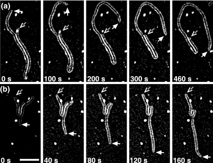
Two time-lapse series of cephalexin-treated cells moving on hard agar stained with FM4-64. (a) First cell, with images taken at 100-s time intervals; (b) second cell, with images taken at 40-s time intervals. Empty arrows indicate the rear ends of the cells; solid white arrows indicate the front ends. Bar on first frame of panel b, 3 μm. The movies are published at our website (http://mcb.berkeley.edu/faculty/BMB/zusmand.html ).
References
-
- Hoiczyk, E., and W. Baumeister. 1998. The junctional pore complex, a prokaryotic secretion organelle, is the molecular motor underlying gliding motility in cyanobacteria. Curr. Biol. 8:1161-1168. - PubMed
-
- Jahn, E. 1924. Beitrage zur botanischen Protistologie. I. Die Polyangiden. Gebruder Borntraeger, Leipzig, Germany.
-
- McBride, M. 2001. Bacterial gliding motility: multiple mechanisms for cell movement over surfaces. Annu. Rev. Microbiol. 55:49-75. - PubMed
Publication types
MeSH terms
Grants and funding
LinkOut - more resources
Full Text Sources
