. 2007 Oct;145(2):527-38.
doi: 10.1104/pp.107.104919. Epub 2007 Aug 17.

Golgi regeneration after brefeldin A treatment in BY-2 cells entails stack enlargement and cisternal growth followed by division

Affiliations

Golgi regeneration after brefeldin A treatment in BY-2 cells entails stack enlargement and cisternal growth followed by division

Markus Langhans et al. Plant Physiol. 2007 Oct.

Abstract

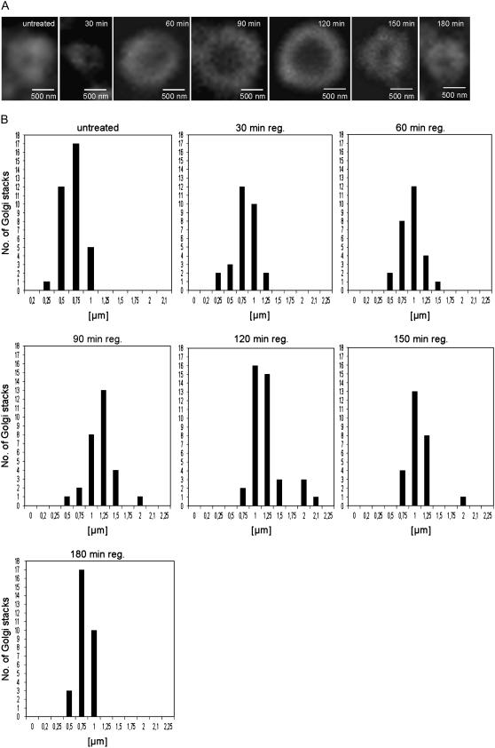
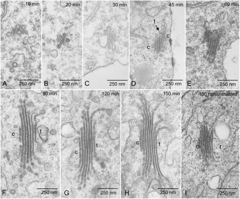
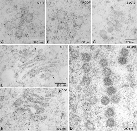
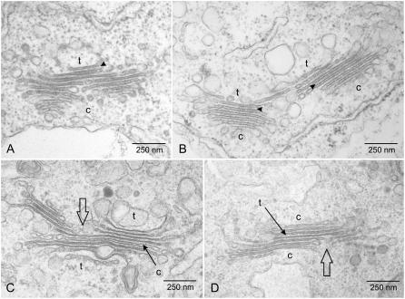
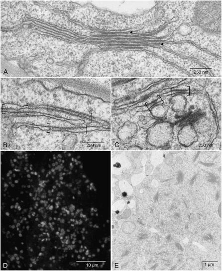
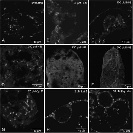
Brefeldin A (BFA) treatment stops secretion and leads to the resorption of much of the Golgi apparatus into the endoplasmic reticulum. This effect is reversible upon washing out the drug, providing a situation for studying Golgi biogenesis. In this investigation Golgi regeneration in synchronized tobacco BY-2 cells was followed by electron microscopy and by the immunofluorescence detection of ARF1, which localizes to the rims of Golgi cisternae and serves as an indicator of COPI vesiculation. Beginning as clusters of vesicles that are COPI positive, mini-Golgi stacks first become recognizable 60 min after BFA washout. They continue to increase in terms of numbers and length of cisternae for a further 90 min before overshooting the size of control Golgi stacks. As a result, increasing numbers of dividing Golgi stacks were observed 120 min after BFA washout. BFA-regeneration experiments performed on cells treated with BFA (10 microg mL(-1)) for only short periods (30-45 min) showed that the formation of ER-Golgi hybrid structures, once initiated by BFA treatment, is an irreversible process, the further incorporation of Golgi membranes into the ER continuing during a subsequent drug washout. Application of the protein kinase A inhibitor H-89, which effectively blocks the reassembly of the Golgi apparatus in mammalian cells, also prevented stack regeneration in BY-2 cells, but only at very high, almost toxic concentrations (>200 microm). Our data suggest that under normal conditions mitosis-related Golgi stack duplication may likely occur via cisternal growth followed by fission.


Figures

Figure 1.
Figure 1.
Golgi stack regeneration during BFA washout in BY-2 cells as monitored by immunofluorescence labeling with ARF1 antibodies. A, Representative individual images from the recovery time points indicated. B, The external diameters of stacks visualized end-on were determined from at least 50 stacks from different cells in samples removed from cultures at the recovery times indicated.
Figure 2.
Figure 2.
Electron microscopy of Golgi stack regeneration. A to I, Representative images of pre-Golgi structures (A and B), mini-Golgis (C–E), and mega-Golgis (F–H) in comparison to a Golgi stack from a wild-type cell not treated with BFA (I). The times at which samples were removed for chemical fixation are given as indicated. Cisternal indicators of polarity were easily recognizable after 90 min of BFA washout but could be occasionally discerned earlier. The Golgi stack presented in D, for instance, is an early example of a cis-trans-cis bipolar stack (see also Fig. 3D). c = cis; t = trans.
Figure 3.
Figure 3.
Immunogold electron microscopy with COPI and COPII antisera. Sections of BY-2 cells prepared by high-pressure freezing/freeze substitution and stained with ARF1, γ-COP for the presence of COPI proteins, and SEC13 for COPII proteins. Samples were removed and freeze-fixed after 20 min (A–D) and 60 min (E and F). c = cis; t = trans.
Figure 4.
Figure 4.
Fission profiles of dividing Golgi stacks found in BY-2 cells 120 to 150 min after BFA washout. In addition to putative cis → trans profiles (A and B), divisions in bipolar stacks were also observed (C and D). c = cis; t = trans.
Figure 5.
Figure 5.
Golgi regeneration after short-term BFA treatment. A, Stacked ER-Golgi hybrid present in wild-type BY-2 cells after 30 min of treatment with 10 μg mL−1 BFA. Intercisternal filaments are visible at different positions throughout the stack. B, Golgi domains in the ER (in brackets) remain recognizable 30 min after BFA washout. C, New Golgi stacks arise although old Golgi domains in the ER are still visible (30 min BFA → 45 min washout). D and E, Massive Golgi stack regeneration after short-term BFA treatment (30 min BFA → 120 min washout; D, confocal image after ARF1 immunostaining; E, chemical-fixed sample.
Figure 6.
Figure 6.
Effects of inhibitors on Golgi stack regeneration. BY-2 cells were treated with BFA (10 μg mL−1) for 45 min, then allowed to regenerate Golgi stacks in the presence or absence of the indicated inhibitor for a 120-min period. Golgi stack presence was monitored by ARF1 immunofluorescence. Images presented are from 45-μm optical sections in the confocal laser scanning microscope. A, Control cells. B to F, Increasing concentrations of H-89. G and H, Microfilament inhibitors cytochalasin D (20 μm) and latrunculin B (2 μm). I, Microtubule inhibitor oryzalin (10 μm).

Similar articles

Cited by

References

    1. Altan-Bonnet N, Phair RD, Polishchuk RS, Weigert R, Lippincott-Schwartz J (2003) A role for Arf1 in mitotic Golgi disassembly, chromosome segregation, and cytokinesis. Proc Natl Acad Sci USA 100 13314–13319 - PMC - PubMed
    1. Altan-Bonnet N, Sougrat R, Lippincott-Schwartz J (2004) Molecular basis for Golgi maintenance and biogenesis. Curr Opin Cell Biol 16 364–372 - PubMed
    1. Altan-Bonnet N, Sougrat R, Liu W, Snapp EL, Ward T, Lippincott-Schwartz J (2006) Golgi inheritance in mammalian cells is mediated through endoplasmic reticulum export activities. Mol Biol Cell 17 990–1005 - PMC - PubMed
    1. Aniento F, Matsuoka K, Robinson DG (2006) ER-to-Golgi transport: the COPII-pathway. In DG Robinson, ed, The Plant Endoplasmic Reticulum. Springer-Verlag, Berlin, pp 99–124
    1. Aridor M, Balch WE (2000) Kinase signaling initiates coat complex II (COPII) recruitment and export from the mammalian endoplasmic reticulum. J Biol Chem 275 35673–35676 - PubMed

Publication types

MeSH terms