microPET of tumor integrin alphavbeta3 expression using 18F-labeled PEGylated tetrameric RGD peptide (18F-FPRGD4)
- PMID: 17704249
- PMCID: PMC4183663
- DOI: 10.2967/jnumed.107.040816
microPET of tumor integrin alphavbeta3 expression using 18F-labeled PEGylated tetrameric RGD peptide (18F-FPRGD4)
Abstract
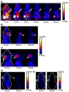
In vivo imaging of alpha(v)beta(3) expression has important diagnostic and therapeutic applications. Multimeric cyclic RGD peptides are capable of improving the integrin alpha(v)beta(3)-binding affinity due to the polyvalency effect. Here we report an example of (18)F-labeled tetrameric RGD peptide for PET of alpha(v)beta(3) expression in both xenograft and spontaneous tumor models.
Methods: The tetrameric RGD peptide E{E[c(RGDyK)](2)}(2) was derived with amino-3,6,9-trioxaundecanoic acid (mini-PEG; PEG is poly(ethylene glycol)) linker through the glutamate alpha-amino group. NH(2)-mini-PEG-E{E[c(RGDyK)](2)}(2) (PRGD4) was labeled with (18)F via the N-succinimidyl-4-(18)F-fluorobenzoate ((18)F-SFB) prosthetic group. The receptor-binding characteristics of the tetrameric RGD peptide tracer (18)F-FPRGD4 were evaluated in vitro by a cell-binding assay and in vivo by quantitative microPET imaging studies.
Results: The decay-corrected radiochemical yield for (18)F-FPRGD4 was about 15%, with a total reaction time of 180 min starting from (18)F-F(-). The PEGylation had minimal effect on integrin-binding affinity of the RGD peptide. (18)F-FPRGD4 has significantly higher tumor uptake compared with monomeric and dimeric RGD peptide tracer analogs. The receptor specificity of (18)F-FPRGD4 in vivo was confirmed by effective blocking of the uptake in both tumors and normal organs or tissues with excess c(RGDyK).
Conclusion: The tetrameric RGD peptide tracer (18)F-FPRGD4 possessing high integrin-binding affinity and favorable biokinetics is a promising tracer for PET of integrin alpha(v)beta(3) expression in cancer and other angiogenesis related diseases.
Figures


Similar articles
-
18F-labeled mini-PEG spacered RGD dimer (18F-FPRGD2): synthesis and microPET imaging of alphavbeta3 integrin expression.Eur J Nucl Med Mol Imaging. 2007 Nov;34(11):1823-1831. doi: 10.1007/s00259-007-0427-0. Epub 2007 May 5. Eur J Nucl Med Mol Imaging. 2007. PMID: 17492285 Free PMC article.
-
Pegylated Arg-Gly-Asp peptide: 64Cu labeling and PET imaging of brain tumor alphavbeta3-integrin expression.J Nucl Med. 2004 Oct;45(10):1776-83. J Nucl Med. 2004. PMID: 15471848
-
[18F]FB-NH-mini-PEG-E{E[c(RGDyK)]2}2.2008 Feb 20 [updated 2008 Mar 12]. In: Molecular Imaging and Contrast Agent Database (MICAD) [Internet]. Bethesda (MD): National Center for Biotechnology Information (US); 2004–2013. 2008 Feb 20 [updated 2008 Mar 12]. In: Molecular Imaging and Contrast Agent Database (MICAD) [Internet]. Bethesda (MD): National Center for Biotechnology Information (US); 2004–2013. PMID: 20641437 Free Books & Documents. Review.
-
18F-labeled galacto and PEGylated RGD dimers for PET imaging of αvβ3 integrin expression.Mol Imaging Biol. 2010 Oct;12(5):530-8. doi: 10.1007/s11307-009-0284-2. Epub 2009 Dec 1. Mol Imaging Biol. 2010. PMID: 19949981 Free PMC article.
-
64Cu-1,4,7,10-Tetraazacyclododecane-N,N',N'',N'''-tetraacetic acid-E{E[c(RGDyK)]2}2.2007 Aug 10 [updated 2007 Aug 29]. In: Molecular Imaging and Contrast Agent Database (MICAD) [Internet]. Bethesda (MD): National Center for Biotechnology Information (US); 2004–2013. 2007 Aug 10 [updated 2007 Aug 29]. In: Molecular Imaging and Contrast Agent Database (MICAD) [Internet]. Bethesda (MD): National Center for Biotechnology Information (US); 2004–2013. PMID: 20641739 Free Books & Documents. Review.
Cited by
-
Radiolabeled Cyclic RGD Peptide Bioconjugates as Radiotracers Targeting Multiple Integrins.Bioconjug Chem. 2015 Aug 19;26(8):1413-38. doi: 10.1021/acs.bioconjchem.5b00327. Epub 2015 Aug 3. Bioconjug Chem. 2015. PMID: 26193072 Free PMC article. Review.
-
Radiolabeled Cyclic RGD Peptides as Radiotracers for Imaging Tumors and Thrombosis by SPECT.Theranostics. 2011 Jan 18;1:58-82. doi: 10.7150/thno/v01p0058. Theranostics. 2011. PMID: 21547153 Free PMC article.
-
Synthesis and Evaluation of a Dimeric RGD Peptide as a Preliminary Study for Radiotheranostics with Radiohalogens.Molecules. 2021 Oct 10;26(20):6107. doi: 10.3390/molecules26206107. Molecules. 2021. PMID: 34684688 Free PMC article.
-
(68)Ga-labeled cyclic RGD dimers with Gly3 and PEG4 linkers: promising agents for tumor integrin alphavbeta3 PET imaging.Eur J Nucl Med Mol Imaging. 2009 Jun;36(6):947-57. doi: 10.1007/s00259-008-1045-1. Epub 2009 Jan 22. Eur J Nucl Med Mol Imaging. 2009. PMID: 19159928
-
In Vivo Characterization of 4 68Ga-Labeled Multimeric RGD Peptides to Image αvβ3 Integrin Expression in 2 Human Tumor Xenograft Mouse Models.J Nucl Med. 2018 Aug;59(8):1296-1301. doi: 10.2967/jnumed.117.206979. Epub 2018 Apr 6. J Nucl Med. 2018. PMID: 29626124 Free PMC article.
References
-
- Castel S, Pagan R, Mitjans F, et al. RGD peptides and monoclonal antibodies, antagonists of αv-integrin, enter the cells by independent endocytic pathways. Lab Invest. 2001;81:1615–1626. - PubMed
-
- Cheng YF, Kramer RH. Human microvascular endothelial cells express integrin-related complexes that mediate adhesion to the extracellular matrix. J Cell Physiol. 1989;139:275–286. - PubMed
-
- Hwang R, Varner J. The role of integrins in tumor angiogenesis. Hematol Oncol Clin North Am. 2004;18:991–1006. - PubMed
-
- Albelda SM, Mette SA, Elder DE, et al. Integrin distribution in malignant melanoma: association of the β3 subunit with tumor progression. Cancer Res. 1990;50:6757–6764. - PubMed
-
- Bello L, Francolini M, Marthyn P, et al. αvβ3 and αvβ5 integrin expression in glioma periphery. Neurosurgery. 2001;49:380–389. discussion 390. - PubMed
Publication types
MeSH terms
Substances
Grants and funding
LinkOut - more resources
Full Text Sources
Other Literature Sources
