. 2007 Dec 1;408(2):241-9.
doi: 10.1042/BJ20070734.

A stress-induced rice (Oryza sativa L.) beta-glucosidase represents a new subfamily of glycosyl hydrolase family 5 containing a fascin-like domain

Affiliations

A stress-induced rice (Oryza sativa L.) beta-glucosidase represents a new subfamily of glycosyl hydrolase family 5 containing a fascin-like domain

Rodjana Opassiri et al. Biochem J. .

Abstract

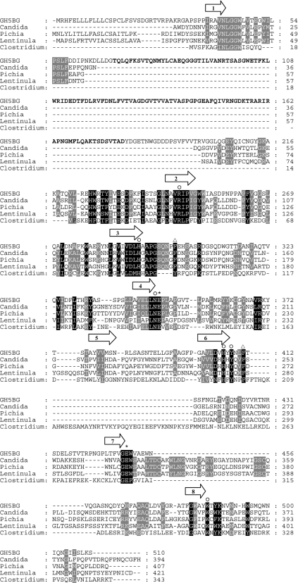
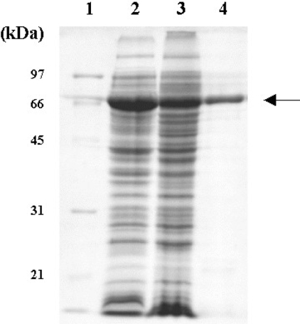
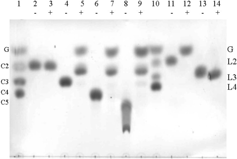
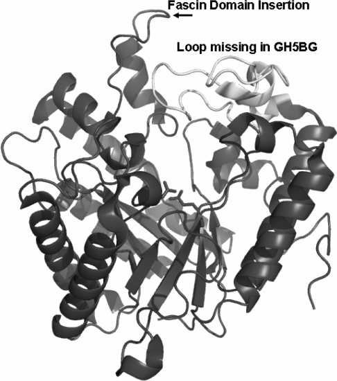
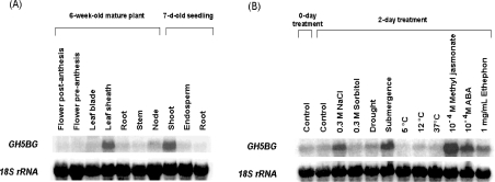
GH5BG, the cDNA for a stress-induced GH5 (glycosyl hydrolase family 5) beta-glucosidase, was cloned from rice (Oryza sativa L.) seedlings. The GH5BG cDNA encodes a 510-amino-acid precursor protein that comprises 19 amino acids of prepeptide and 491 amino acids of mature protein. The protein was predicted to be extracellular. The mature protein is a member of a plant-specific subgroup of the GH5 exoglucanase subfamily that contains two major domains, a beta-1,3-exoglucanase-like domain and a fascin-like domain that is not commonly found in plant enzymes. The GH5BG mRNA is highly expressed in the shoot during germination and in leaf sheaths of mature plants. The GH5BG was up-regulated in response to salt stress, submergence stress, methyl jasmonate and abscisic acid in rice seedlings. A GUS (glucuronidase) reporter tagged at the C-terminus of GH5BG was found to be secreted to the apoplast when expressed in onion (Allium cepa) cells. A thioredoxin fusion protein produced from the GH5BG cDNA in Escherichia coli hydrolysed various pNP (p-nitrophenyl) glycosides, including beta-D-glucoside, alpha-L-arabinoside, beta-D-fucoside, beta-D-galactoside, beta-D-xyloside and beta-D-cellobioside, as well as beta-(1,4)-linked glucose oligosaccharides and beta-(1,3)-linked disaccharide (laminaribiose). The catalytic efficiency (kcat/K(m)) for hydrolysis of beta-(1,4)-linked oligosaccharides by the enzyme remained constant as the DP (degree of polymerization) increased from 3 to 5. This substrate specificity is significantly different from fungal GH5 exoglucanases, such as the exo-beta-(1,3)-glucanase of the yeast Candida albicans, which may correlate with a marked reduction in a loop that makes up the active-site wall in the Candida enzyme.


Figures

Figure 1
Figure 1. Alignment of the protein sequence of rice GH5BG with exo-β-1,3-glucanases and endo-β-1,4-glucanase
GH5BG is rice GH5BG, Candida is exo-β-(1,3)-glucanase from Candida albicans (AC CAA39908), Lentinula is exo-β-(1,3)-glucanase from Lentinula edodes (AC AB192344), Pichia is exo-β-(1,3)-glucanase from Pichia pastoris (AC AY954499), and Clostridium is endo-β-(1,4)-glucanase from Clostridium thermocellum (AC AAA23220). The alignment was generated with the ClustalX implementation of ClustalW [27,28] and analysed and manually adjusted with Gendoc [29]. Alignment of the C. thermocellum sequence relied on the structural alignment of the 1CEC structural model with the C. albicans Exg 1CZ1 structure. The positions of the β-strands of the central (β/α)8 barrel are indicated by arrows above the alignment. The asterisks identify the two catalytic glutamate residues, the invariant GH family 5 residues are marked by the symbol ○ above the column, and the black and grey shadings highlight other identities between sequences. The two phenylalanine residues found at the +1 subsite of C. albicans Exg are marked by triangles above the column. The region of rice GH5BG homologous with fascin is indicated by bold text.
Figure 2
Figure 2. SDS/PAGE of GH5BG–thioredoxin fusion protein expressed in E. coli strain Origami B (DE3) after incubation in the presence of 0.5 mM IPTG at 20 °C for 12 h
Lanes: 1, standard marker (Bio-Rad); 2, total protein of E. coli cells containing pET32a(+)/DEST-GH5BG; 3, soluble fraction of E. coli cells containing pET32a(+)/DEST-GH5BG; 4, purified thioredoxin–GH5BG. The arrow points to the thioredoxin–GH5BG.
Figure 3
Figure 3. Hydrolysis of disaccharides and oligosaccharide substrates by thioredoxin–GH5BG detected by TLC
The thioredoxin–GH5BG was incubated with 5 mM substrates for 30 min and the products were detected by the carbohydrate staining method described in the Experimental section. Samples were incubated with (+) or without (−) enzyme in 50 mM sodium acetate, pH 5.0, for 30 min at 37 °C prior to being spotted on silica-gel 60 F254 TLC plates, developed and charred with 10% H2SO4 in ethyl alcohol. Lanes: 1, glucose (G) and cello-oligosaccharides of DP 2–4 (C2–C4) marker; 2 and 3, cellobiose; 4 and 5, cellotriose; 6 and 7, cellotetraose; 8 and 9, cellopentaose; 10, standard laminari-oligosaccharides of DP 2–4 (L2–L4); 11 and 12, laminaribiose; 13 and 14, laminaritriose.
Figure 4
Figure 4. Active site of Candida albicans Exg structural model with differences in the loops around the active site found in rice GH5BG highlighted
The 1CZ1 structure [9] is shown as a ribbon diagram coloured dark grey, with the loop after β-strand 7 of the (β/α)8 barrel shown in white and labelled to draw attention to its absence in rice GH5BG. The insertion of the fascin-like domain after the first helix of the extended loop after strand 1 of the β-barrel is indicated by the label. The catalytic acid/base (left) and catalytic nucleophile (right) are displayed in stick representation to indicate the location of the active site. The image was generated with Pymol [30].
Figure 5
Figure 5. Northern-blot analysis of GH5BG transcript levels in (A) 7-day-old rice seedlings and 6-week-old mature plant tissues and (B) 7-day-old rice seedlings grown a further 2 days with various abiotic stresses and plant hormones
GH5BG, RNA blots were probed with α-32P-labelled GH5BG gene-specific probe; 18S rRNA indicates the same blot probed with an α-32P-labelled 18 S rRNA cDNA probe. A 20 μg portion of total RNA from the appropriate tissues was loaded in each lane.
Figure 6
Figure 6. Extracellular localization of the GH5BG protein
The epidermal layers of onion bulbs were transformed with (A) pMDC139-GH5BG and (B) control pMDC139 plasmid without insert by particle bombardment. After 48 h incubation at 25 °C in complete darkness, the transformed onion cells were stained overnight with X-Glu at 37 °C, and the location of blue-coloured product was observed by optical microscopy.

References

    1. Henrissat B., Davies G. T. Structural and sequence-based classification of glycoside hydrolases. Curr. Opin. Struct. Biol. 1997;7:637–644. - PubMed
    1. Jenkins J., Leggio L. L., Harris G., Pickersgill R. β-Glucosidase, β-galactosidase, family A cellulases, family F xylanases and two barley glycanases form a superfamily of enzymes with 8-fold β/α architecture and with two conserved glutamates near the carboxy-terminal ends of β-strands four and seven. FEBS Lett. 1995;362:281–285. - PubMed
    1. Henrissat B., Callebaut I., Fabrega S., Lehn P., Mornon J.-P., Davies G. Conserved catalytic machinery and the prediction of a common fold for several families of glycosyl hydrolases. Proc. Natl. Acad. Sci. U.S.A. 1995;92:7090–7094. - PMC - PubMed
    1. Coutinho P. M., Henrissat B. Carbohydrate-active enzymes: an integrated database approach. In: Gilbert H. J., Davies G., Henrissat B., Svensson B., editors. Recent Advances in Carbohydrate Bioengineering. Cambridge: Royal Society of Chemistry; 1999. pp. 3–12.
    1. Gilkes N. R., Henrissat B., Kilburn D. G., Miller R. C., Jr, Warren R. A. J. Domains in microbial β-1,4-glycanases: sequence conservation, function, and enzyme families. Microbiol. Rev. 1991;55:303–315. - PMC - PubMed

Publication types

MeSH terms