. 2007 Aug 16:7:96.
doi: 10.1186/1471-213X-7-96.

Regionalization of the mouse visceral endoderm as the blastocyst transforms into the egg cylinder

Affiliations

Regionalization of the mouse visceral endoderm as the blastocyst transforms into the egg cylinder

Aitana Perea-Gomez et al. BMC Dev Biol. .

Abstract

Background: Reciprocal interactions between two extra-embryonic tissues, the extra-embryonic ectoderm and the visceral endoderm, and the pluripotent epiblast, are required for the establishment of anterior-posterior polarity in the mouse. After implantation, two visceral endoderm cell types can be distinguished, in the embryonic and extra-embryonic regions of the egg cylinder. In the embryonic region, the specification of the anterior visceral endoderm (AVE) is central to the process of anterior-posterior patterning. Despite recent advances in our understanding of the molecular interactions underlying the differentiation of the visceral endoderm, little is known about how cells colonise the three regions of the tissue.

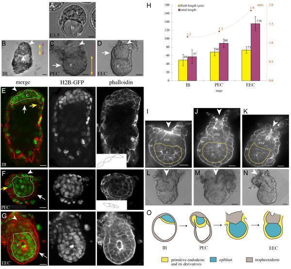
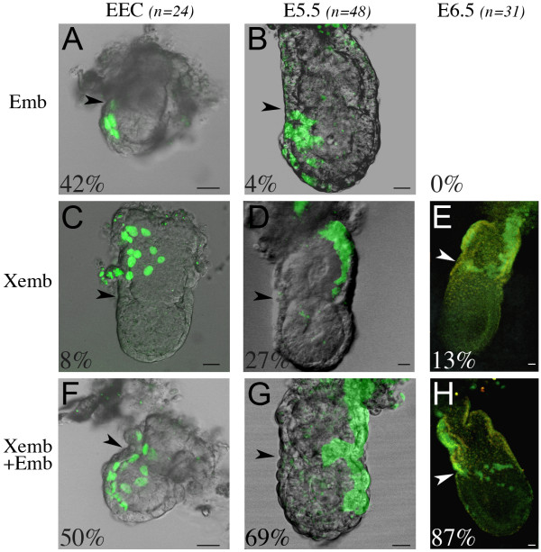
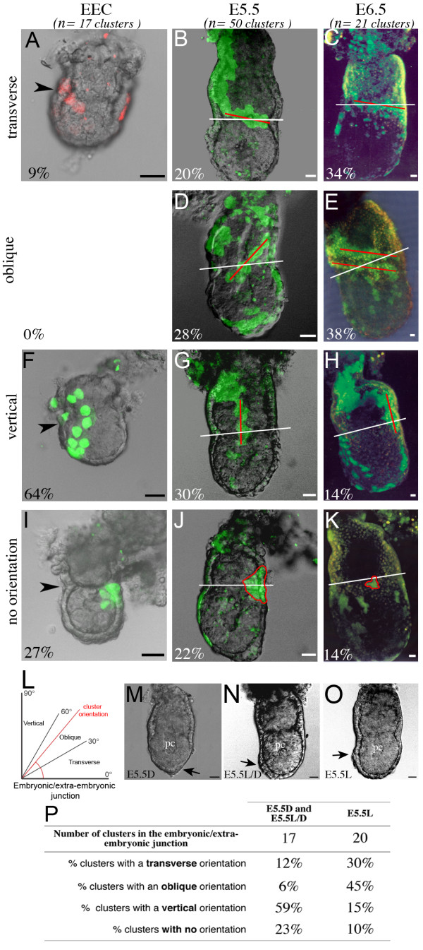
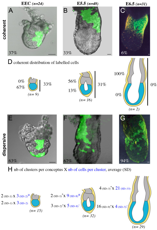
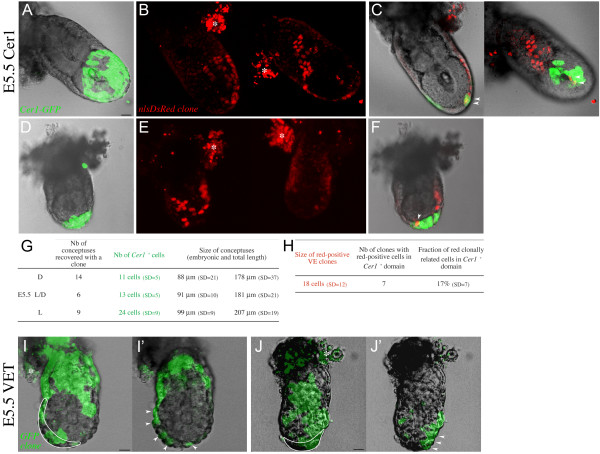
Results: As a first step, we performed morphological observations to understand how the extra-embryonic region of the egg cylinder forms from the blastocyst. Our analysis suggests a new model for the formation of this region involving cell rearrangements such as folding of the extra-embryonic ectoderm at the early egg cylinder stage. To trace visceral endoderm cells, we microinjected mRNAs encoding fluorescent proteins into single surface cells of the inner cell mass of the blastocyst and analysed the distribution of labelled cells at E5.0, E5.5 and E6.5. We found that at E5.0 the embryonic and extra-embryonic regions of the visceral endoderm do not correspond to distinct cellular compartments. Clusters of labelled cells may span the junction between the two regions even after the appearance of histological and molecular differences at E5.5. We show that in the embryonic region cell dispersion increases after the migration of the AVE. At this time, visceral endoderm cell clusters tend to become oriented parallel to the junction between the embryonic and extra-embryonic regions. Finally we investigated the origin of the AVE and demonstrated that this anterior signalling centre arises from more than a single precursor between E3.5 and E5.5.

Conclusion: We propose a new model for the formation of the extra-embryonic region of the egg cylinder involving a folding of the extra-embryonic ectoderm. Our analyses of the pattern of labelled visceral endoderm cells indicate that distinct cell behaviour in the embryonic and extra-embryonic regions is most apparent upon AVE migration. We also demonstrate the polyclonal origin of the AVE. Taken together, these studies lead to further insights into the formation of the extra-embryonic tissues as they first develop after implantation.


Figures

Figure 1
Figure 1
Formation of the extra-embryonic region of the egg cylinder. (A-D) Bright field images of mouse conceptuses, before implantation at E3.5 (A), and after implantation at E4.7-E5.0 (B-D). Arrowheads point to the polar trophectoderm, later called extra-embryonic ectoderm (from pre-egg cylinder stage). White arrows show the visceral endoderm. b, blastocoel ; e, epiblast ; EEC, early egg cylinder; i, inner cell mass (ICM) ; IB, implanted blastocyst ; m, mural trophectoderm; PEC, pre-egg cylinder. (E-G) Phalloidin-Texas red staining (red) of H2B-GFP (green) transgenic conceptuses, showing respectively the membranes and nuclei of cells. Confocal sections of conceptuses at the implanted blastocyst (IB in E), pre-egg cylinder (PEC in F) and early egg cylinder (EEC in G) stages are shown. Yellow arrows show the parietal endoderm. The contour of the epiblast is highlighted in white. Insets show the contour of 3 neighbouring cells of the polar trophectoderm. (H) Measurements of the embryonic region (Emb) and total length of conceptuses at E4.7-E5.0, as defined by the coloured double arrows in B-C. The average number is shown and error bars indicate the standard deviation from 12 implanted blastocysts, 18 pre-egg cylinder and 24 early egg cylinder conceptuses. The ratio between the two lengths is represented in brown. pc, proamniotic cavity. Conceptuses are oriented with the proximal and distal poles respectively up and down. (I-K) Confocal sections of E4.8-E5.0 conceptuses stained with phalloidin-Texas red, showing the distribution of actin. Arrowheads point to an enrichment of actin in a folded region of extra-embryonic ectoderm (exe). The contour of the epiblast is highlighted in yellow. ve, visceral endoderm. (L-N) Bright field images of E4.8-E5.0 conceptuses. Arrowheads point to the folding of the polar trophectoderm. (O) Schematic summary showing the characteristics of conceptuses ordered by stage during the formation of the extra-embryonic region. The polar trophectoderm (grey) thickens and folds, whereas the primitive endoderm (yellow), which initially covers the epiblast (implanted blastocyst stage) and mural trophectoderm (pre-egg cylinder stage), engulfs the extra-embryonic ectoderm at the early egg cylinder stage. Scale bars 20 μm.
Figure 2
Figure 2
Lack of growth restriction between the embryonic and extra-embryonic regions of the VE. Examples of labelled conceptuses recovered after microinjection of single cells at the surface of the ICM at the blastocyst stage. They are shown as a fluorescent projection of a confocal z series merged with a transmitted light section, at the early egg cylinder (EEC) stage (A, C, F), E5.5 (B, D, G) and E6.5 (E, H). For each stage, the number of labelled conceptuses considered (n) and the percentage of those with colonisation of the embryonic region only (Emb, A-B), the extra-embryonic region only (Xemb, C-E) or both (Xemb+Emb, F-H) is indicated. Arrowheads point to the embryonic/extra-embryonic junction. Conceptuses are oriented with the anterior pole on the left, whenever it can be identified. Scale bars 20 μm.
Figure 3
Figure 3
Shift in orientation of VE clusters at the embryonic/extra-embryonic junction. (A-K) Examples, shown as a fluorescent projection of a confocal z series merged with a transmitted light section, of VE clusters with a transverse orientation (A-C), oblique orientation (D-E), vertical orientation (F-H) and with no specific orientation (I-K) at the level of the embryonic/extra-embryonic junction at the early egg cylinder (EEC) stage (A, F, I), E5.5 (B, D, G, J) and E6.5 (C, E, H, K). The orientation of clusters at E5.5 and E6.5 is indicated by a red line and that of the embryonic/extra-embryonic junction by a white line. Arrowheads indicate the embryonic/extra-embryonic junction at the early egg cylinder stage. The contour of clusters with no specific orientation is outlined by a red line. For each stage, the number of clusters considered (n) and the percentage of clusters with a given orientation are shown. The number of conceptuses with clusters of labelled cells at the embryonic/extra-embryonic junction considered is 17 at the early egg cylinder stage, 36 at E5.5 and 21 at E6.5. Some conceptuses have several clusters at the embryonic/extra-embryonic junction. (L) Schematic representation of the classification of junction clusters according to the value of the angle between the orientation of the cluster (red line) and that of the embryonic/extra-embryonic junction. (M-O) Bright field images of conceptuses at E5.5. Black arrows show the thickening of the visceral endoderm (VET), which indicates the position of the anterior pole, distally (D), laterodistally (L/D) or laterally (L). (P) Summary table of the orientation of junction clusters at E5.5 according to the position of the VET. Scale bars 20 μm.
Figure 4
Figure 4
Cell dispersion in the VE of the embryonic region specifically increases after E5.5. Examples, shown as a fluorescent projection of a confocal z series merged with a transmitted light section, of coherent (A-C) and dispersive (E-G) distributions of labelled cells at the early egg cylinder (EEC) stage (A, E), E5.5 (B, F) and E6.5 (C, G). For each stage, the number of conceptuses considered and the percentage of coherent and dispersive cases are indicated. (D) Schematic representation of the localisation of labelled cells in coherent distributions. The number of conceptuses considered at each stage is indicated (n) and the percentage of cases with labelled cells in the extra-embryonic region (grey), the embryonic region (blue) or spanning both (vertical bar) is shown. (H) Schematic representation of the characteristics of dispersive distributions of labelled cells in the extra-embryonic region (grey) or embryonic regions (blue). The number of conceptuses considered at each stage (n) is indicated. The average and the standard deviation of the number of clusters per conceptus are shown in black. The number of conceptuses considered with clusters in the extra-embryonic and embryonic regions respectively is 9 and 12 at the early egg cylinder stage, 20 and 21 at E5.5, 26 and 27 at E6.5. Some conceptuses have both extra-embryonic and embryonic clusters. The average and the standard deviation of the number of cells per clusters are shown in dark blue. The number of clusters considered in the extra-embryonic and embryonic regions respectively is 14 and 21 at the early egg cylinder stage, 40 and 53 at E5.5, 92 and 431 at E6.5. *,°,+,~ are significantly different (Mann-Whitney test, p < 0.05). nb, number; SD standard deviation. Scale bars 20 μm.
Figure 5
Figure 5
Polyclonal origin of the Anterior Visceral Endoderm (AVE) region. (A-H) The Cer1-GFP transgenic line is used as a marker of the AVE region. Examples of two conceptuses (A-C and D-F) at E5.5 containing a red-positive clone in the VE. (A, D) Cer1-GFP pattern indicates the embryonic stage, respectively E5.5L and E5.5D. Green fluorescent projection of a confocal z series merged with a transmitted light section are shown. (B, E) VE clones labelled with a red nuclear marker. The two sides of the conceptus are shown as a red fluorescent projection of a confocal z series. Asterisks indicate autofluorescence of the parietal endoderm. (C, F) Merged confocal sections of the conceptuses, showing the colocalisation (arrowheads) of the transgenic green and clonal red markers. (G) Summary of the characteristics of conceptuses with a given Cer1-GFP pattern. Average numbers are given. D, distal ; L/D, laterodistal; L, lateral; nb, number. (H) Summary of the characteristics of red-positive clones in the transgenic line. Average numbers are given. VE, visceral endoderm. (I-J) The visceral endoderm thickening (VET) is used as a marker of the AVE region and is delineated by a white line. Examples of GFP-positive cells in wild type conceptuses, at E5.5L/D in I and E5.5D in J. Arrowheads point to the positive cells colonising the VET. I, J are green fluorescent projection of a confocal z series merged with a transmitted light section and I', J' are merged confocal sections. Scale bars 20 μm.

References

    1. Beddington RS, Robertson EJ. Axis development and early asymmetry in mammals. Cell. 1999;96:195–209. doi: 10.1016/S0092-8674(00)80560-7. - DOI - PubMed
    1. Ang SL, Constam DB. A gene network establishing polarity in the early mouse embryo. Semin Cell Dev Biol. 2004;15:555–561. doi: 10.1016/j.semcdb.2004.04.009. - DOI - PubMed
    1. Copp AJ. Interaction between inner cell mass and trophectoderm of the mouse blastocyst. II. The fate of the polar trophectoderm. J Embryol Exp Morphol. 1979;51:109–120. - PubMed
    1. Batten BE, Haar JL. Fine structural differentiation of germ layers in the mouse at the time of mesoderm formation. Anat Rec. 1979;194:125–141. doi: 10.1002/ar.1091940109. - DOI - PubMed
    1. Rivera-Perez JA, Mager J, Magnuson T. Dynamic morphogenetic events characterize the mouse visceral endoderm. Dev Biol. 2003;261:470–487. doi: 10.1016/S0012-1606(03)00302-6. - DOI - PubMed

Publication types