

Cell-to-cell spread and massive vacuole formation after Cryptococcus neoformans infection of murine macrophages
- PMID: 17705844
- PMCID: PMC1988836
- DOI: 10.1186/1471-2172-8-16
Cell-to-cell spread and massive vacuole formation after Cryptococcus neoformans infection of murine macrophages
Abstract
Background: The interaction between macrophages and Cryptococcus neoformans (Cn) is critical for containing dissemination of this pathogenic yeast. However, Cn can either lyse macrophages or escape from within them through a process known as phagosomal extrusion. Both events result in live extracellular yeasts capable of reproducing and disseminating in the extracellular milieu. Another method of exiting the intracellular confines of cells is through host cell-to-cell transfer of the pathogen, and this commonly occurs with the human immuno-deficiency virus (HIV) and CD4+ T cells and macrophages. In this report we have used time-lapse imaging to determine if this occurs with Cn.
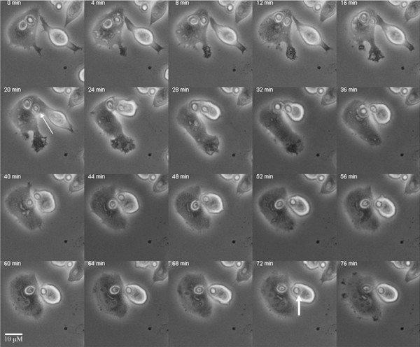
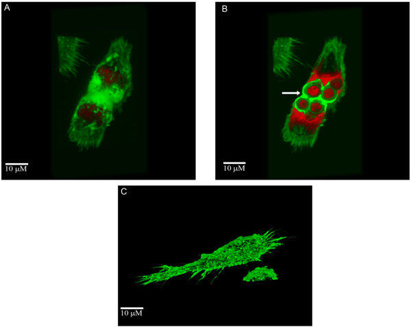
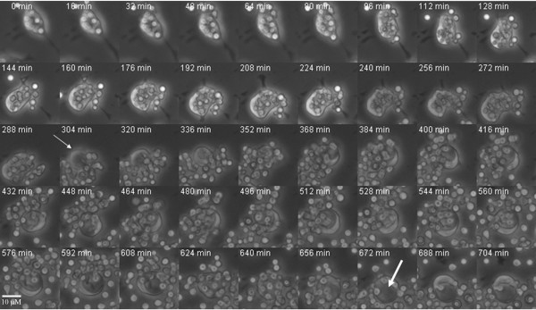
Results: Live imaging of Cryptococcus neoformans interactions with murine macrophages revealed cell-to-cell spread of yeast cells from infected donor cells to uninfected cells. Although this phenomenon was relatively rare its occurrence documents a new capacity for this pathogen to infect adjacent cells without exiting the intracellular space. Cell-to-cell spread appeared to be an actin-dependent process. In addition, we noted that cryptococcal phagosomal extrusion was followed by the formation of massive vacuoles suggesting that intracellular residence is accompanied by long lasting damage to host cells.
Conclusion: C. neoformans can escape the intracellular confines of macrophages in an actin dependent manner by cell-to-cell transfer of the yeast leading to infection of adjacent cells. In addition, complete extrusion of internalized Cn cells can lead to the formation of a massive vacuole which may be a sign of damage to the host macrophage. These observations document new outcomes for the interaction of C. neoformans with host cells that provide precedents for cell biological effects that may contribute to the pathogenesis of cryptococcal infections.
Figures
References
-
- Shao X, Mednick A, Alvarez M, van Rooijen N, Casadevall A, Goldman DL. An innate immune system cell is a major determinant of species-related susceptibility differences to fungal pneumonia. J Immunol. 2005;175:3244–3251. - PubMed
-
- Lee SC, Kress Y, Zhao ML, Dickson DW, Casadevall A. Cryptococcus neoformans survive and replicate in human microglia. Lab Invest. 1995;73:871–879. - PubMed
Publication types
MeSH terms
Grants and funding
LinkOut - more resources
Full Text Sources
Other Literature Sources
Research Materials
