. 2007 Aug 17:7:140.
doi: 10.1186/1471-2148-7-140.

Multiple domain insertions and losses in the evolution of the Rab prenylation complex

Affiliations

Multiple domain insertions and losses in the evolution of the Rab prenylation complex

Rita Rasteiro et al. BMC Evol Biol. .

Abstract

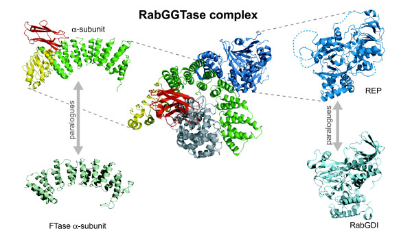
Background: Rab proteins are regulators of vesicular trafficking, requiring a lipid modification for proper function, prenylation of C-terminal cysteines. This is catalysed by a complex of a catalytic heterodimer (Rab Geranylgeranyl Transferase - RabGGTase) and an accessory protein (Rab Escort Protein. REP). Components of this complex display domain insertions relative to paralogous proteins. The function of these inserted domains is unclear.

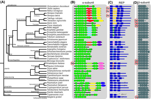
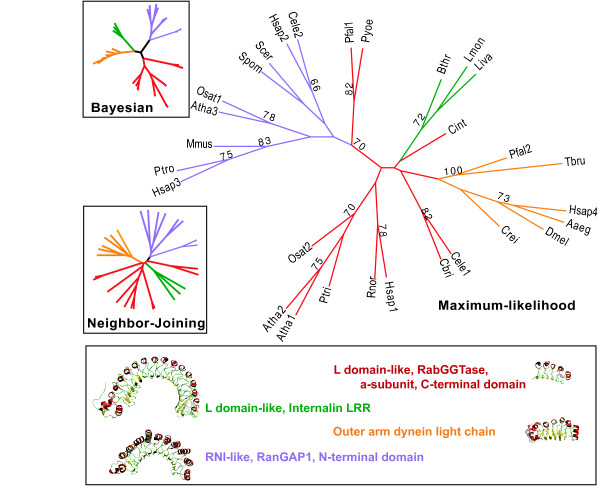
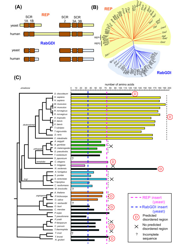
Results: We profiled the domain architecture of the components of the Rab prenylation complex in evolution. We identified the orthologues of the components of the Rab prenylation machinery in 43 organisms, representing the crown eukaryotic groups. We characterize in detail the domain structure of all these components and the phylogenetic relationships between the individual domains.

Conclusion: We found different domain insertions in different taxa, in alpha-subunits of RGGTase and REP. Our results suggest that there were multiple insertions, expansions and contractions in the evolution of this prenylation complex.


Figures

Figure 1
Figure 1
Cartoon representing the three dimensional structure of Rat RabGGTase complexed with REP-1 (1LTX) [29]. The α subunit is shown in green with a yellow and red parts, representing a Leucine Rich Repeat and a C2-like domain, respectively. REP1 is shown in blue with dotted blue lines representing disordered regions. The β subunit is shown in gray. The α-subunit and REP are shown in isolation on the left and right side respectively, next to cartoons representing the three dimensional structure of the paralogous α-subunit of FTase and of RabGDI (PDB codes 1FT1 [11] and 1LV0 [72], respectively).
Figure 2
Figure 2
(A) taxonomic tree of the organisms analyzed in this study, adapted from the NCBI's taxonomy database [73] and from reference [28] (B) N- to C-terminal domain organization of the α-subunit of RGGTase. Boxes with the same shape represent the same domain, whereas different shapes represent distinct domains. Green boxes represent tetratricopeptide repeats (TPRs). In between the 5th and 6th TPR some proteins display an inserted domain. In deuterostomes this is a C2-like domain (inverted orange triangle), but in other organisms the inserted sequences are not recognized as any known domain. They are represented by colored lines. Inserts of the same color are similar to each other. Ellipses represent LRR domains – distinct colors represent sequence similarity. The red circle with 2× on the left indicates that this protein is duplicated in that organism. (C) N- to C-terminal domain organization of REP. The blue boxes represent the Sequence Conserved Regions (SCRs) between SCR1 and SCR2 there is an inserted domain of unknown structure. This domain is only conserved within taxa: all the inserted sequences represented in yellow are similar to each other but distinct from all other inserted sequences represented in other colors. (D) N- to C-terminal domain organization of the β-subunit The gray boxes represent the prenyltransferase and squalene oxidase repeat.
Figure 3
Figure 3
Maximum Likelihood reconstruction of the evolutionary relationships between the LRR domains found in RGGTase a-subunit (red branches) and reference sequences representing other superfamilies of LRR domains – L-domain-like, internalin LRR (106v) – green; RNI-like, 28-residue LRR, Ribonucelase inhibitor (1A4Y) – blue; RNI-like, RanGAP1, N-terminal domain(1K5D) – purple; outer arm dynein light chain(1M9L) – orange; L-domain-like, RabGGTase α-subunit, C-terminal domain (1LTX) – red. Inset boxes show the topologies of Bayesian and Neighbor Joining trees of the same sequences, showing contradicting topologies. Species codes are Aaeg – Aedes aegypti; Atha – Arabidopsis thaliana; Bthr-Bacillus thrugiensis; Cbri – Caenorhabditis briggsae; Cele – Caenorhabditis elegans; Cint – Ciona intestinalis; Crei – Chlamydomonas reinhardtii; Dmel – Drosophila melanogaster; Hsap – Homo sapiens; Liva – Listeria ivanovii, Lmon – Listeria monocytogenes; Mmus – Mus musculus; Osat – Oryza sativa; Pfal-Plasmodium falciparum; Ptri – Populus trichocarpa; Ptro – Pan troglodytes; Pyoe-Plamodium yoelii; Rnor – Rattus norvegicus; Scer – Saccharomyces cerevisae;Spom – Schizosaccharomyces pombe; Tbru – Trypanosoma brucei
Figure 4
Figure 4
(A) scheme illustrating the location of the sequence conserved regions (SCRs) in RabGDI and REP sequences, as well as the variable size of the region between SCR1A and SCR2. The broken yellow and blue boxes indicate the "gap" in the alignment of these sequences with mammalian REP. Adapted from [13] (B) Neighbor-Joining tree of selected sequences of REP and RabGDI, illustrating that classification of these sequences into one of the two subfamilies is clear. The same tree with bootstrap vaues is provided as supplementary Figure S6. Species codes are Aaeg – Aedes aegypti; Afum – Aspergillus fumigatus; Agam – Anopheles gambiae; Atha – Arabidopsis thaliana; Cbri – Caenorhabditis briggsae; Cele-Caenorhabditis elegans; Cint – Ciona intestinalis; Cmer – Cyanidioschyzon merolae; Cneo – Cryptococcus neoformans; Cpar – Cryptosporodium parvum; Crei-Chlamydomonas reinhardtii; Ddis – Dictyostelium discoideum; Dmel – Drosophila melanogaster; Dpse – Drosophila pseudoobscura; Drer – Danio rerio; Gint-Giardis intestinalis; Hsap – Homo sapiens; Mbre – Monosiga brevicollis; Mmus-Mus musculus; Ngru – Naegleria gruberi; Nvec – Nematostella vectensis; Osat-Oryza sativa; Otau – Ostreococcus tauri; Pber – Plasmodium berghei; Pfal – Plasmodium falciparum; Ppyg-Pongo pygmaeus; Pram – Phytophthora ramorum; Psoj – Phytophthora sojae; Ptri-Populus trichocarpa; Pyoe – Plamodium yoelii; Rnor – Rattus norvegicus; Scer-Saccharomyces cerevisae; Sjap – Schistosoma japonicum; Spom-Schizosaccharomyces pombe; Tbru – Trypanosoma brucei; Tcas – Tribolium castaneum; Tcru – Trypanosoma cruzi; Tpse-Thalassiosira pseudonana; Tnig – Tetraodon nigroviridis; Trub – Takifugu rubripes; Tthe – Tetrahymena thermophila; Xlae – Xenopus laevis; Xtro – Xenopus tropicalis; Ylip – Yarrowia lipolytica (C) Plot of the size of the region between SCR1A and SCR2 in the different species, measured in number of amino acids. The bars are coded according to the colors used in Figure 2 and same color denotes identifiable sequence similarity. The blue and pink dotted perpendicular lines are for reference and indicate the number of amino acids between SCR1B and SCR2 for RabGDI and MRS6, respectively. A red D indicates that the insert contains a predicted disordered region [51], whereas a cross means that such regions are not predicted in the insert.

Similar articles

Cited by

References

    1. Maurer-Stroh S, Washietl S, Eisenhaber F. Protein prenyltransferases. Genome Biol. 2003;4:212. doi: 10.1186/gb-2003-4-4-212. - DOI - PMC - PubMed
    1. Casey PJ, Seabra MC. Protein prenyltransferases. J Biol Chem. 1996;271:5289–5292. doi: 10.1074/jbc.271.10.5289. - DOI - PubMed
    1. Lane KT, Beese LS. Thematic review series: lipid posttranslational modifications. Structural biology of protein farnesyltransferase and geranylgeranyltransferase type I. J Lipid Res. 2006;47:681–699. doi: 10.1194/jlr.R600002-JLR200. - DOI - PubMed
    1. Leung KF, Baron R, Seabra MC. Thematic review series: lipid posttranslational modifications. geranylgeranylation of Rab GTPases. J Lipid Res. 2006;47:467–475. doi: 10.1194/jlr.R500017-JLR200. - DOI - PubMed
    1. McTaggart SJ. Isoprenylated proteins. Cell Mol Life Sci. 2006;63:255–267. doi: 10.1007/s00018-005-5298-6. - DOI - PMC - PubMed

Substances

LinkOut - more resources