

Non-canonical Wnt signaling enhances differentiation of Sca1+/c-kit+ adipose-derived murine stromal vascular cells into spontaneously beating cardiac myocytes
- PMID: 17706246
- PMCID: PMC2048991
- DOI: 10.1016/j.yjmcc.2007.06.012
Non-canonical Wnt signaling enhances differentiation of Sca1+/c-kit+ adipose-derived murine stromal vascular cells into spontaneously beating cardiac myocytes
Abstract
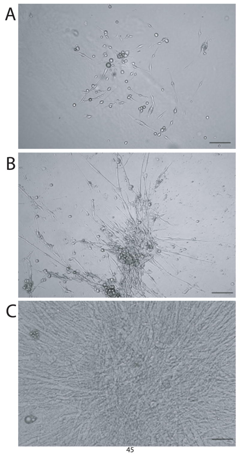
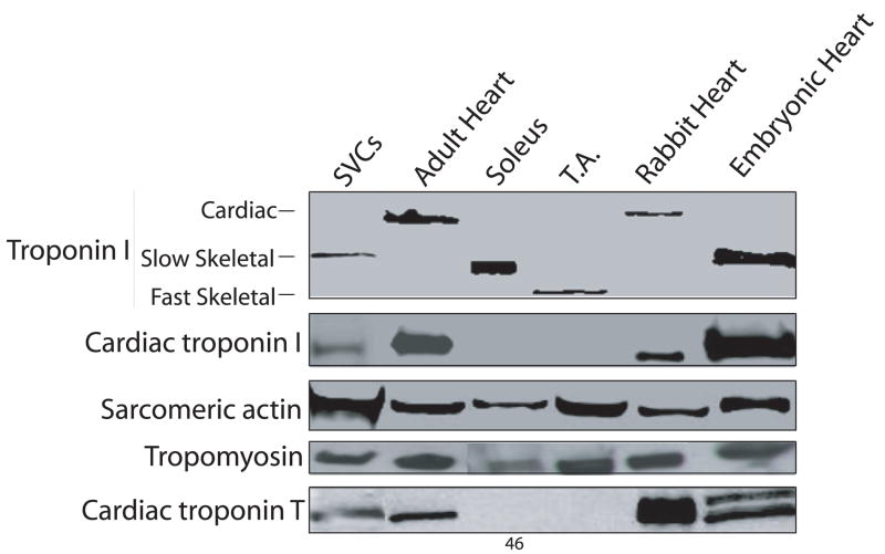
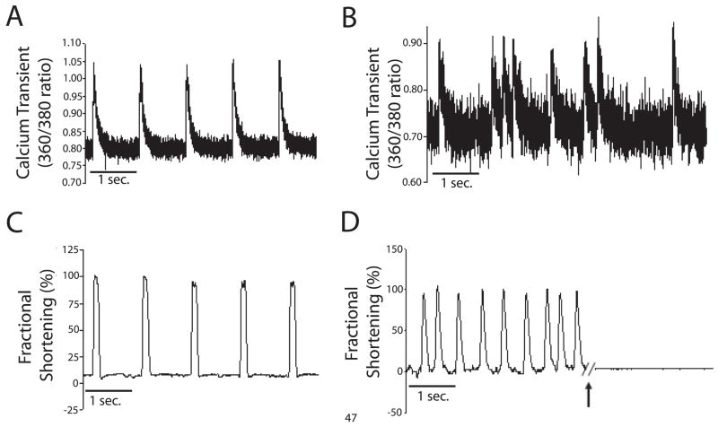
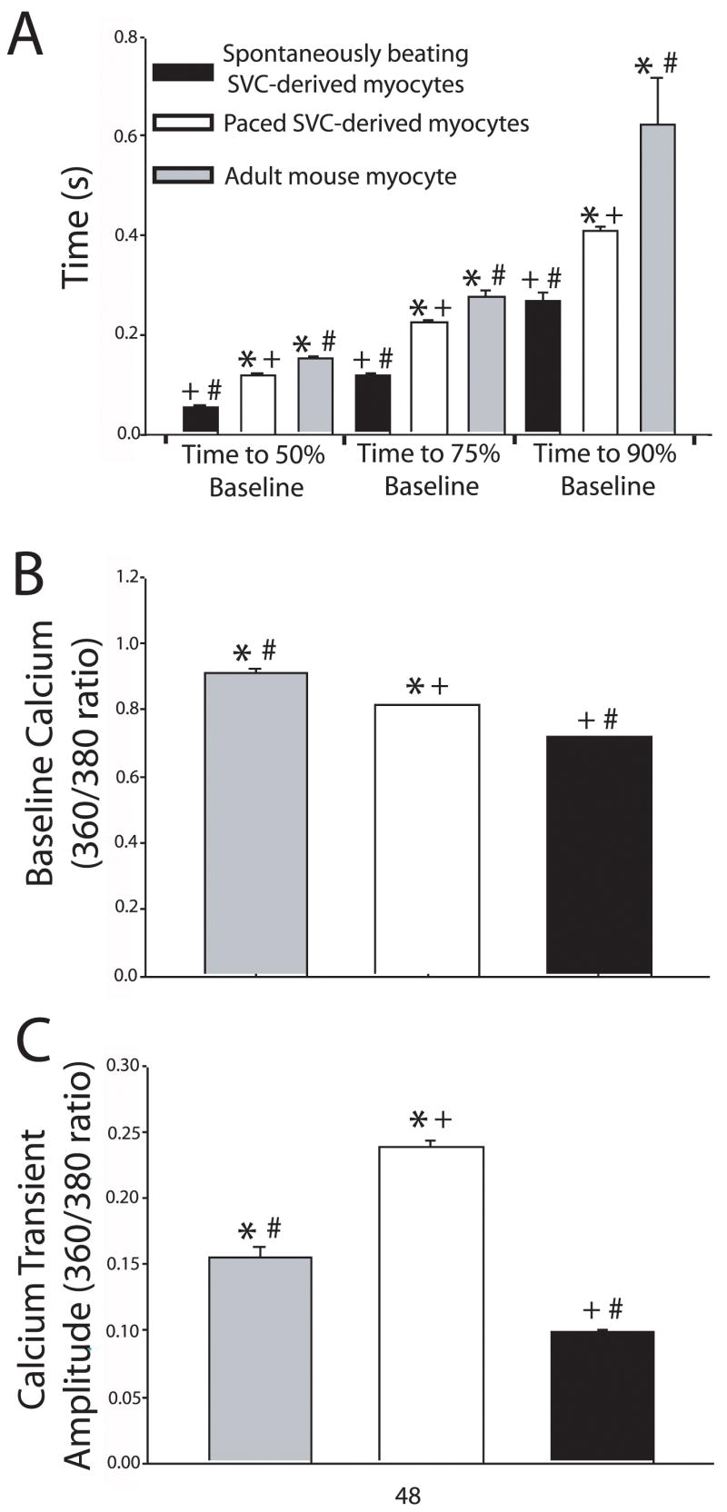
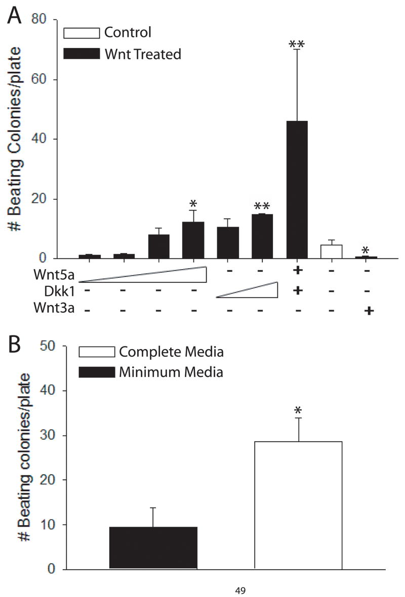
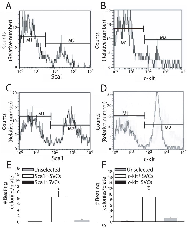
Recent reports have described a stem cell population termed stromal vascular cells (SVCs) derived from the stromal vascular fraction of adipose tissue, which are capable of intrinsic differentiation into spontaneously beating cardiomyocytes in vitro. The objective of this study was to further define the cardiac lineage differentiation potential of SVCs in vitro and to establish methods for enriching SVC-derived beating cardiac myocytes. SVCs were isolated from the stromal vascular fraction of murine adipose tissue. Cells were cultured in methylcellulose-based murine stem cell media. Analysis of SVC-derived beating myocytes included Western blot and calcium imaging. Enrichment of acutely isolated SVCs was carried out using antibody-tagged magnetic nanoparticles, and pharmacologic manipulation of Wnt and cytokine signaling. Under initial media conditions, spontaneously beating SVCs expressed both cardiac developmental and adult protein isoforms. Functionally, this specialized population can spontaneously contract and pace under field stimulation and shows the presence of coordinated calcium transients. Importantly, this study provides evidence for two independent mechanisms of enriching the cardiac differentiation of SVCs. First, this study shows that differentiation of SVCs into cardiac myocytes is augmented by non-canonical Wnt agonists, canonical Wnt antagonists, and cytokines. Second, SVCs capable of cardiac lineage differentiation can be enriched by selection for stem cell-specific membrane markers Sca1 and c-kit. Adipose-derived SVCs are a unique population of stem cells that show evidence of cardiac lineage development making them a potential source for stem cell-based cardiac regeneration studies.
Figures
Similar articles
-
Clonal multilineage differentiation of murine common pluripotent stem cells isolated from skeletal muscle and adipose stromal cells.Ann N Y Acad Sci. 2005 Jun;1044:183-200. doi: 10.1196/annals.1349.024. Ann N Y Acad Sci. 2005. PMID: 15958712
-
Myocyte-specific enhancer factor 2c triggers transdifferentiation of adipose tissue-derived stromal cells into spontaneously beating cardiomyocyte-like cells.Sci Rep. 2021 Jan 15;11(1):1520. doi: 10.1038/s41598-020-80848-3. Sci Rep. 2021. PMID: 33452355 Free PMC article.
-
Identification of osteo-adipo progenitor cells in fat tissue.Cell Prolif. 2008 Oct;41(5):803-12. doi: 10.1111/j.1365-2184.2008.00542.x. Cell Prolif. 2008. PMID: 18616697 Free PMC article.
-
The potential of adipose-derived adult stem cells as a source of neuronal progenitor cells.Plast Reconstr Surg. 2005 Oct;116(5):1453-60. doi: 10.1097/01.prs.0000182570.62814.e3. Plast Reconstr Surg. 2005. PMID: 16217495 Review.
-
Revisiting the Advances in Isolation, Characterization and Secretome of Adipose-Derived Stromal/Stem Cells.Int J Mol Sci. 2018 Jul 27;19(8):2200. doi: 10.3390/ijms19082200. Int J Mol Sci. 2018. PMID: 30060511 Free PMC article. Review.
Cited by
-
Pathogenic peptide deviations support a model of adaptive evolution of chordate cardiac performance by troponin mutations.Physiol Genomics. 2010 Jul 7;42(2):287-99. doi: 10.1152/physiolgenomics.00033.2010. Epub 2010 Apr 27. Physiol Genomics. 2010. PMID: 20423961 Free PMC article.
-
Hypoxic culture and in vivo inflammatory environments affect the assumption of pericyte characteristics by human adipose and bone marrow progenitor cells.Am J Physiol Cell Physiol. 2011 Dec;301(6):C1378-88. doi: 10.1152/ajpcell.00460.2010. Epub 2011 Aug 24. Am J Physiol Cell Physiol. 2011. PMID: 21865587 Free PMC article.
-
Biomaterial Approaches for Stem Cell-Based Myocardial Tissue Engineering.Biomark Insights. 2015 Jun 1;10(Suppl 1):77-90. doi: 10.4137/BMI.S20313. eCollection 2015. Biomark Insights. 2015. PMID: 26052226 Free PMC article. Review.
-
Stromal vascular fraction transplantation as an alternative therapy for ischemic heart failure: anti-inflammatory role.J Cardiothorac Surg. 2011 Mar 31;6:43. doi: 10.1186/1749-8090-6-43. J Cardiothorac Surg. 2011. PMID: 21453457 Free PMC article.
-
Stem cell factor gene transfer promotes cardiac repair after myocardial infarction via in situ recruitment and expansion of c-kit+ cells.Circ Res. 2012 Nov 9;111(11):1434-45. doi: 10.1161/CIRCRESAHA.111.263830. Epub 2012 Aug 29. Circ Res. 2012. PMID: 22931954 Free PMC article.
References
-
- Bai X, Pinkernell K, Song YH, Nabzdyk C, Reiser J, Alt E. Genetically selected stem cells from human adipose tissue express cardiac markers. Biochem Biophys Res Commun. 2007;353:665–671. - PubMed
-
- Bunting KD, Hawley RG. Integrative molecular and developmental biology of adult stem cells. Biology of the Cell. 2003;95:563–578. - PubMed
-
- Cannon RO. Cardiovascular potential of BM-derived stem and progenitor cells. Cytotherapy. 2004;6:602–607. - PubMed
-
- Choi SC, Yoon J, Shim WJ, Ro YM, Lim DS. 5-azacytidine induces cardiac differentiation of P19 embryonic stem cells. Experimental and Molecular Medicine. 2004;36:515–523. - PubMed
-
- Day SM, Westfall MV, Fomicheva EV, Hoyer K, Yasuda S, La Cross NC, D’Alecy LG, Ingwall JS, Metzger JM. Histidine button engineered into cardiac troponin I protects the ischemic and failing heart. Nat Med. 2006;12:181–189. - PubMed
Publication types
MeSH terms
Substances
Grants and funding
LinkOut - more resources
Full Text Sources
Other Literature Sources
