. 2007 Sep 21;372(3):608-18.
doi: 10.1016/j.jmb.2007.05.084. Epub 2007 Jun 2.

Molecular basis of tropomyosin binding to tropomodulin, an actin-capping protein

Affiliations

Molecular basis of tropomyosin binding to tropomodulin, an actin-capping protein

Alla S Kostyukova et al. J Mol Biol. .

Abstract

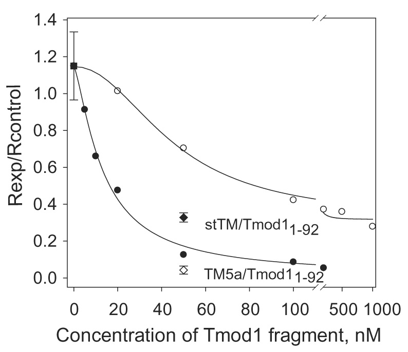
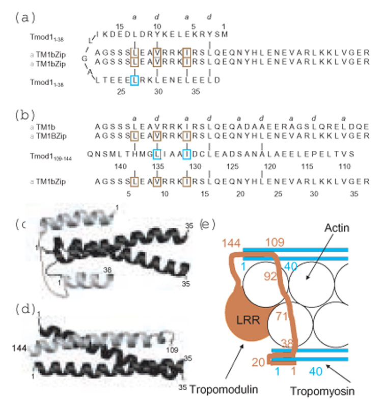
The tropomodulin (Tmod) family of proteins that cap the pointed, slow-growing end of actin filaments require tropomyosin (TM) for optimal function. Earlier studies identified two regions in Tmod1 that bind the N terminus of TM, though the ability of different isoforms to bind the two sites is controversial. We used model peptides to determine the affinity and define the specificity of the highly conserved N termini of three short, non-muscle TMs (alpha, gamma, delta-TM) for the two Tmod1 binding sites using circular dichroism spectroscopy, native gel electrophoresis, and chemical crosslinking. All TM peptides have high affinity for the second Tmod1 binding site (within residues 109-144; alpha-TM, 2.5 nM; gamma-TM, delta-TM, 40-90 nM), but differ >100-fold for the first site (residues 1-38; alpha-TM, 90 nM; undetectable at 10 microM, gamma-TM, delta-TM). Residue 14 (R in alpha; Q in gamma and delta) and, to a lesser extent, residue 4 (S in alpha; T in gamma and delta) are primarily responsible for the differences. The functional consequence of the sequence differences is reflected in more effective inhibition of actin filament elongation by full-length alpha-TMs than gamma-TM in the presence of Tmod1. The binding sites of the two Tmod1 peptides on a model TM peptide differ, as defined by comparing (15)N,(1)H HSQC spectra of a (15)N-labeled model TM peptide in both the absence and presence of Tmod1 peptide. The NMR and CD studies show that there is an increase in alpha-helix upon Tmod1-TM complex formation, indicating that intrinsically disordered regions of the two proteins become ordered upon binding. A model proposed for the binding of Tmod to actin and TM at the pointed end of the filament shows how the Tmod-TM accentuates the asymmetry of the pointed end and suggests how subtle differences among TM isoforms may modulate actin filament dynamics.


Figures

Figure 1
Figure 1
Complex formation between Tmod1 and TM peptides monitored by non-denaturing polyacrylamide gel-electrophoresis. Lane 1 and 4, Tmod1; lane 2, Tmod1 and αTM1bZip; lane 3, Tmod1 and γTM1bZip. TM peptides are positively charged and do not enter the gel. Arrowhead indicates Tmod1, arrows indicate complexes.
Figure 2
Figure 2
Binding of Tmod1 fragments to TM peptides measured using circular dichroism spectroscopy. (A): Tmod1109–144 and γTM1bZip, the peptide concentrations were 10 µM in 100 mM NaCl, 10 mM sodium phosphate, pH 7.0. (B): Tmod11–38 and γTM1bZip, and (C): Tmod11–38 and δTM1bZip, the peptide concentrations were 30 µM in 100 mM NaCl, 10 mM sodium phosphate, pH 7.0. (●) Tmod1 peptide; (○)TM peptide; (▽) sum of the folding curves of TM and Tmod1 peptides; (▼) the folding of the mixture of TM and Tmod1 peptides.
Figure 3
Figure 3
The cross-linking of TM model peptides and Tmod1 N-terminal fragments with glutaraldehyde monitored by the SDS-PAGE (15 % polyacrylamide). A: Complex formation: lane 1, Tmod11–38; lane 2, Tmod11–38 treated with glutaraldehyde; lane 3, Tmod11–38/ αTM1bZip; lane 4, Tmod11–38/ αTM1bZip treated with glutaraldehyde; lane 5, Tmod11–38/ γTM1bZip; lane 6, Tmod11–38/ γTM1bZip treated with glutaraldehyde; lane 7, Tmod11–92 treated with glutaraldehyde; lane 8, Tmod11–92/ γTM1bZip; lane 9, Tmod11–92/ γTM1bZip treated with glutaraldehyde; lane 10, Tmod11–92. B: Control for non-specific cross-linking: lane 1, TM9d252–284; lane 2, TM9d252–284 treated with glutaraldehyde; lane 3, Tmod11–92/ TM9d252–284 treated with glutaraldehyde; lane 4, γTM1bZip treated with glutaraldehyde. The arrowheads indicate individual proteins, whereas the arrows indicate complexes.
Figure 4
Figure 4
Dependence of inhibition of pointed end elongation of gelsolin-capped actin filaments on Tmod11–344 or Tmod11–92 concentration in the presence of 2.5 µM γTM5NM1. Initial rates (R) were calculated as the first derivatives at time zero after fitting exponential growth curves to the data. The inhibition of polymerization was calculated as Rexp / Rcontrol, where Rcontrol=1 (in the absence of tropomyosin and tropomodulin). (●) with Tmod11–344; (○) with Tmod11–92. (■) the value shown for γTM5NM1 in the absence of tropomodulin is 1.15 ± 0.18 (n=3). For comparison initial rates in the presence of 50 nM Tmod11–92 with 0.75 µM αstTM (◆) or 0.5 µM αTM5a (◇) are shown.
Figure 5
Figure 5
Complex formation between Tmod11–38 and TM peptides monitored by non-denaturing polyacrylamide gel-electrophoresis. Lane 1, Tmod11–38; lane 2, Tmod11–38 and αTM1bZip; lane 3, Tmod11–38 and γTM1bZip; lane 4, Tmod11–38 and δTM1bZip; lane 5, Tmod11–38 and αTM1bZip(S4T); lane 6, Tmod11–38 and αTM1bZip(R148Q); lane 7, Tmod11–38 and αTM1bZip (S4T/R148Q); lane 8, Tmod11–38 and αTM1bZip(S15V/E18Q). TM peptides are positively charged and do not enter the gel. The difference in mobility of complexes is due to isoelectric points of TM peptides (pI=10.4 for αTM1bZip(S15V/E18Q) and 9.9 for αTM1bZip and αTM1bZip(S4T). Arrow indicates Tmod11–38, arrowhead indicates complexes.
Figure 6
Figure 6
15N-¹H HSQC spectra of 15N-αTM1bZip in the presence and absence of tropomodulin fragments. Panel A, (red) 15N-αTM1bZip, (cyan) 15N-αTM1bZip + unlabeled Tmod11–38. Panel B, (red) 15N- αTM1bZip, (cyan) 15N-αTM1bZip + unlabeled Tmod1109–144. The peptides were each ~0.5 mM in 100 mM NaCl, 10 mM NaCl, 5% deuterium oxide, pH 6.5. Data were acquired on a Bruker 800 MHz NMR spectrometer. Note in unbound αTM1bZip Q19 overlaps R11 and V27 overlaps N20.
Figure 7
Figure 7
Schematic of possible binding modes of Tmod11–38 and Tmod1109–144 to αTM1bZip. (a, b) The sequences and possible coiled-coil interactions at a and d positions of the heptad repeat of the two Tmod binding sites for αTM1bZip. Mutations in Tmod1 that cause loss of binding to TM ; ; ; are outlined in cyan, and mutations in TM5NM1 that cause loss of binding to Tmod1 are outlined in brown . (c,d) Models of possible structures of the binding interfaces. In the models, the two chains of αTm1bZip are black and the single chains of the two Tmod1 peptides are grey. (a,c) Residues 1–13 of Tmod11–38 bind antiparallel to the αTM1bZip coiled coil on one side of the tropomyosin interface, residues 15 to 26 loop around the N-terminus of TM and residues 24–36 bind parallel to the other side of the tropomyosin coiled coil interface. (b,d) Residues 121–138 of Tmod1109–144 bind antiparallel to residues 6 to 23 of αTm1bZip to form a three helix bundle. (e) Cartoon model of the binding of Tmod (terra cotta) to tropomyosin (cyan) at the pointed end of the actin filament (white) to illustrate how one Tmod1 may interact with the N-termini of two TM molecules on opposite sides of the actin helix to cap the pointed end. The TM-dependent capping site includes residue 71 and is shown binding to actin. The TM-independent capping site at the extreme C-terminus of Tmod1 is not shown.

Similar articles

Cited by

References

    1. Pollard TD, Borisy GG. Cellular motility driven by assembly and disassembly of actin filaments. Cell. 2003;112:453–465. - PubMed
    1. Fowler VM. Regulation of actin filament length in erythrocytes and striated muscle. Curr Opin Cell Biol. 1996;8:86–96. - PubMed
    1. Littlefield R, Almenar-Queralt A, Fowler VM. Actin dynamics at pointed ends regulates thin filament length in striated muscle. Nat Cell Biol. 2001;3:544–551. - PubMed
    1. Gunning PW, Schevzov G, Kee AJ, Hardeman EC. Tropomyosin isoforms: divining rods for actin cytoskeleton function. Trends Cell Biol. 2005;15:333–341. - PubMed
    1. Greenfield NJ, Fowler VM. Tropomyosin requires an intact N-terminal coiled coil to interact with tropomodulin. Biophys J. 2002;82:2580–2591. - PMC - PubMed

Publication types

MeSH terms

LinkOut - more resources