. 2007 Nov;156(1):24-31.
doi: 10.1016/j.molbiopara.2007.06.010. Epub 2007 Jun 28.

JBP2, a SWI2/SNF2-like protein, regulates de novo telomeric DNA glycosylation in bloodstream form Trypanosoma brucei

Affiliations

JBP2, a SWI2/SNF2-like protein, regulates de novo telomeric DNA glycosylation in bloodstream form Trypanosoma brucei

Rudo Kieft et al. Mol Biochem Parasitol. 2007 Nov.

Abstract

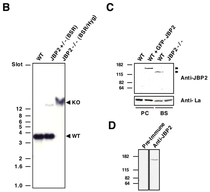
Synthesis of the modified thymine base, beta-d-glucosyl-hydroxymethyluracil or J, within telomeric DNA of Trypanosoma brucei correlates with the bloodstream form specific epigenetic silencing of telomeric variant surface glycoprotein genes involved in antigenic variation. In order to analyze the function of base J in the regulation of antigenic variation, we are characterizing the regulatory mechanism of J biosynthesis. We have recently proposed a model in which chromatin remodeling by a SWI2/SNF2-like protein (JBP2) regulates the developmental and de novo site-specific localization of J synthesis within bloodstream form trypanosome DNA. Consistent with this model, we now show that JBP2 (-/-) bloodstream form trypanosomes contain five-fold less base J and are unable to stimulate de novo J synthesis in newly generated telomeric arrays.


Figures

Figure 1
Figure 1. JBP2 is involved in J-Biosynthesis in Bloodstream Form Trypanosomes
A. Restriction enzyme map of the JBP2 gene locus, with the constructs used to disrupt JBP2 expression shown below. In each case, the JBP2 ORF is replaced by an expression cassette giving either Blasticidin (BSR) or hygromycin (Hyg) resistance. All ORF’s are indicated with grey boxes. Both resistance genes are flanked by α-tubulin intergenic regions (hatched boxes) for proper processing. Restriction enzyme sites used for making the constructs and for mapping of the transformants are indicated: EcoRV (E), HindIII (H), NarI (N). The probe fragment is shown above the map. The 3.6 kbp WT and 20 kbp KO hybridizing EcoRV fragments are indicated (dotted line). B. Southern mapping of bloodstream form T. brucei cell line 221a of strain 427 transformed with the JBP2 knockout constructs. Genomic DNA from wildtype cells (WT), heterozygous (JBP2 +/− (BSR)) and homozygous (JBP2 −/− (BSR/Hyg)) JBP2 knockout cells was digested with EcoRV and hybridized with a JBP2 upstream and ORF probe. Size markers are in indicated in kbp. C. JBP2 is absent in JBP2 −/− bloodstream form trypanosomes. Total cell extracts of the following cell lines were analyzed by Western blotting using anti-JBP2 antisera (α-JBP2): Procyclic 29–13 cells (PC WT), Procyclic WT cells + GFP-JBP2 (PC WT + JBP2), Bloodstream form 221a WT cells (BS WT) and Bloodstream form JBP2 −/− cells (BS JBP2 −/−). The 120 kDa wild type JBP2 protein and the 150 kDa GFP-JBP2 fusion protein are indicated, as well as the position of the size markers (in kDa). The blot was subsequently hybridized with anti-La antisera (α-La) to control for protein loading. D. Specificity of the JBP2 antisera. Total cell extract from procyclic cells expressing the GFP-JBP2 fusion was analyzed by western blot using pre-immune serum (PIM) or anti-JBP2 antisera (Anti-JBP2) at the same dilution. E. J levels in the DNA of JBP2 knockout cells. The J content was determined with anti-J DNA immunoblots. Dot blots with twofold dilution series of DNA from Bloodstream form 221a WT cells (WT), heterozygous JBP2 knockout cells (JBP2 +/− (BSR)), homozygous JBP2 knockout cells (JBP2 −/− (BSR/Hyg)) and homozygous JBP2 knockout cells (JBP2 −/− (BSR/Hyg)) with the ectopically expressed JBP2-GFP were incubated with anti-J antisera. Procyclic (PC) DNA was included as a negative control. Bound antibodies were detected by a second antibody conjugated to horseradish peroxidase and visualized by ECL (left panel). DNA loading was checked by hybridization with a probe specific for the β-Tubulin gene (right panel). F. Localization of J in the JBP2 knockout cell line. DNA from the indicated cell lines was sonicated and immunoprecipitated with anti-J antisera and blotted onto nitrocellulose. The blots were then hybridized with a radiolabeled DNA probe corresponding to the indicated regions of the genome.
Figure 2
Figure 2. JBP2 Stimulates De Novo J-Synthesis
A. Generation of new telomeric arrays in the 221 VSG expression site by Telomere Fragmentation. As described previously [12], transfection of T. brucei with pTMF replaces the native telomere at the 221 bloodstream form expression site (ES) with a new telomere seed 5 kbp downstream of the ribosomal RNA promoter (PrRNA). The question mark refers to the unknown presence of J within newly generated telomeric arrays, both in WT and in JBP2 −/− cells transfected with the pTMF construct. The plasmid was digested with SmaI (M) prior to transfection. Restriction enzyme sites used for mapping of the transformants are indicated: XhoI (X), Bsp120I (B) and ScaI (S). The probe fragments are shown underneath the map. B. Growth of newly generated telomeres in WT and JBP2 −/− cells. From WT cells DNA was isolated 15 and 40 days post transfection; from JBP2 −/− cells 18 and 60 days post transfection. DNA was digested with Xho I and analyzed by Southern blotting. Similar hybridization patterns with the NEO and 221 VSG probe confirm proper integration of the construct in the 221 ES C. JBP2 stimulates de novo J-synthesis in newly generated telomeric DNA. All indicated cell lines were transfected with the pTMF construct. Clones were obtained after which genomic DNA from transfected wildtype cells (WT), transfected homozygous JBP2 knockout cells (JBP2 −/−), transfected homozygous JBP2 knockout cells (JBP2 −/−) grown in the presence of 1 mM HOMedU (HMU) and transfected homozygous JBP2 knockout cells (JBP2 −/−) with the ectopically expressed JBP2-GFP, was isolated and digested with XhoI. An immunoprecipitation was performed with anti-J antisera, after which 10% of the input (In) DNA and 100% of the immunoprecipitated (IP) DNA was analyzed by hybridization with a NEO probe. D. IP efficiency in the JBP2 −/−. DNA was isolated from TMF transfected wild type (WT) and JBP2 knockout (J2KO) and analyzed by immunoprecipitation as described in C. The DNA samples were then dot-blotted onto nitrocellulose and hybridized with the NEO probe (left) and 177bp probe (right)., E. J levels in the DNA of JBP2 knockout cells. The qualitative assessment of J content of the cell lines described in panel C was determined with anti-J DNA immunoblots and ECL detection, as described in Fig. 1 E.

References

    1. Gommers-Ampt JH, Van Leeuwen F, de Beer AL, et al. beta-D-glucosyl-hydroxymethyluracil: a novel modified base present in the DNA of the parasitic protozoan T. brucei. Cell. 1993;75(6):1129–36. - PubMed
    1. Borst P, van Leeuwen F. beta-D-glucosyl-hydroxymethyluracil, a novel base in African trypanosomes and other Kinetoplastida. Molecular & Biochemical Parasitology. 1997;90(1):1–8. - PubMed
    1. van Leeuwen F, Taylor MC, Mondragon A, Moreau H, Gibson W, Kieft R, Borst P. beta-D-glucosyl-hydroxymethyluracil is a conserved DNA modification in kinetoplastid protozoans and is abundant in their telomeres. Proceedings of the National Academy of Sciences of the United States of America. 1998;95(5):2366–71. [see comments] - PMC - PubMed
    1. van Leeuwen F, Wijsman ER, Kuyl-Yeheskiely E, van der Marel GA, van Boom JH, Borst P. The telomeric GGGTTA repeats of Trypanosoma brucei contain the hypermodified base J in both strands. Nucleic Acids Research. 1996;24(13):2476–82. - PMC - PubMed
    1. van Leeuwen F, Wijsman ER, Kieft R, van der Marel GA, van Boom JH, Borst P. Localization of the modified base J in telomeric VSG gene expression sites of Trypanosoma brucei. Genes & Development. 1997;11(23):3232–41. - PMC - PubMed

Publication types

MeSH terms