Comparative Study
. 2007 Oct;90(4):482-92.
doi: 10.1016/j.ygeno.2007.06.007. Epub 2007 Aug 15.

Promoter, alternative splice forms, and genomic structure of protocadherin 15

Affiliations
Comparative Study

Promoter, alternative splice forms, and genomic structure of protocadherin 15

Kumar N Alagramam et al. Genomics. 2007 Oct.

Abstract

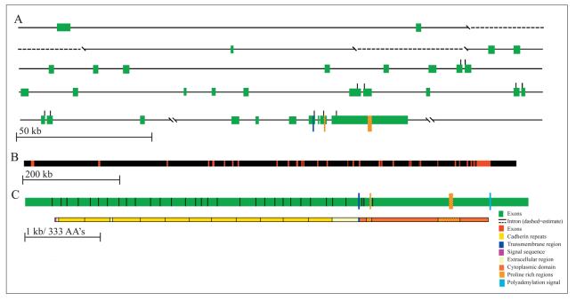
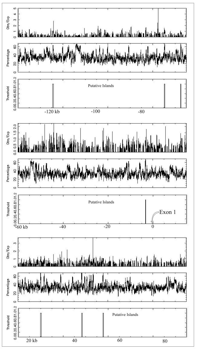
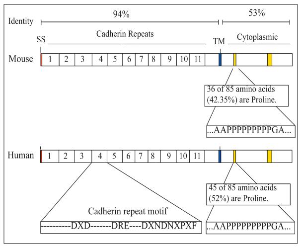
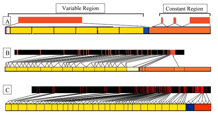
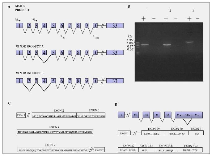
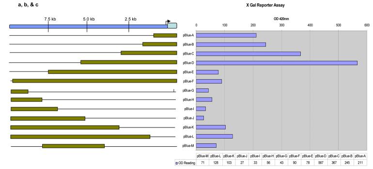
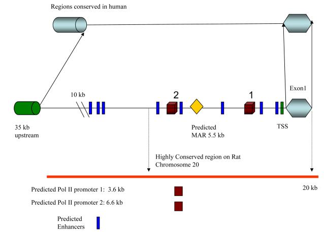
We originally showed that the protocadherin 15 gene (Pcdh15) is necessary for hearing and balance functions; mutations in Pcdh15 affect hair cell development in Ames waltzer (av) mice. Here we extend that study to understand better how Pcdh15 operates in a cell. The original report identified 33 exons in Pcdh15, with exon 1 being noncoding; additional exons of Pcdh15 have since been reported. The 33 exons of Pcdh15 described originally are embedded in 409 kb of mouse genomic sequence, while the corresponding exons of human PCDH15 are spread over 980 kb of genomic DNA; the exons in Pcdh15/PCDH15 range in size from 9 to approximately 2000 bp. The genomic organization of Pcdh15/PCDH15 bears similarity to that of cadherin 23, but differs significantly from other protocadherin genes, such as Pcdhalpha, beta, or gamma. A CpG island is located approximately 2900 bp upstream of the PCDH15 transcriptional start site. The Pcdh15/PCDH15 promoter lacks TATAA or CAAT sequences within 100 bases upstream of the transcription start site; deletion mapping showed that Pcdh15 harbors suppressor and enhancer elements. Preliminary searches for alternatively spliced transcripts of Pcdh15 identified novel splice variants not reported previously. Results from our study show that both mouse and human protocadherin 15 genes have complex genomic structures and transcription control mechanisms.


Figures

Figure 1
Figure 1
Genomic Structure of human PCDH15. A. Green boxes represent individual exons 1-33. (proportional to other exons, but not to scale of diagram) Dotted line indicates gaps where genomic sequence could not be aligned, but sizes could be inferred from contig NT_024078.5. B. Scaled down figure of the PCDH15 gene to show all exons in a single line and their positions along the ∼981 kb genomic DNA. C. Spliced exons aligned with predicted amino acid sequence and predicted domains.
Figure 2
Figure 2
CpG Island: 225 kb of human PCDH15 genomic sequence analyzed for CG content and putative CpG islands. The test thresholds for a reported island were an observed/expected ratio 0.60, a percent C + percent G > 40.00, and a length >200 bp. The putative island directly upstream of exon 1 has a length of 215 bp.
Figure 3
Figure 3
Comparison of similarities in predicted amino acid sequence between mouse and human. TM = transmembrane domain. SS = Signal Sequence
Figure 4
Figure 4
Comparison of the genomic organization of A.)Pcdh alpha/gamma clusters to B.) Pcdh15 to C.) Cdh23. In A the upper orange bars represent the coding genomic regions. The lower bar represents the generic protein structure of members of the Pcdh alpha/gamma clusters. Notice that one large region of DNA codes for all of the cadherin repeats in the cytoplasmic domain (yellow). In B and C the upper orange and black bars represent coding genomic regions. Exons are the orange hash marks and the black regions represent introns. The lower bars for B and C represent the protein structure for Pcdh15 and Cdh23 respectively. Notice that the cadherin repeats in both Cdh23 and Pcdh15 are coded for by multiple exons.
Figure 5
Figure 5
Analysis of the various transcripts produced by alternative splicing in Pcdh15. A. Alternative spliced forms of Pcdh15. This schematic represents (not to scale) the exons in the Protocadherin 15 gene with forward and reverse primers (94,95,108,109) used in thePCR reaction. The major and minor products are shown with all included exons attached by solid lines. Note: The numbering of the exons in this figure is based on the original description of Pcdh15 [3; 4]. To use the criteria established by Haywood-Watson et. al.[15] one exon should come before the first exon shown and all exon numbers in the figure would increase by one. B. Gel analysis of RT-PCR products This PCR reaction includes phi markers (PhiX174) on left and in lane (1) primer sets 94 & 95, in lane (2) primer sets 108 & 109, and in lane (3) primer sets 94 & 109. Both the major and minor alternatively spliced products are present in this PCR reaction. C. Amino acid sequence of exons 2, 3, 4 and 5 of Pchd15. D. Various Transcripts produced by alternative splicing within exon 33.(Upper) Represents 3′ end of Protocadherin 15 gene with minor expressed protein product shown which splices out 33.b segment. (Lower) Represents segment of exons present in 3′ region with amino acid sequences included. Bold face amino acid sequence represent sequence alternatively spliced out in the minor product of this gene.
Figure 6
Figure 6
A. Diagram showing the various fragments used for transfection and reporter assays. The encompassed region includes exon 1 and 10kb upstream. The plasmids ( pBlue TOPO ® vector from invitrogen) (lettered A- M) used contain a lacZ reporter gene downstream of the above fragments. B. Shows the results of transfection and reporter assay of the fragments shown in figure A. Numerical data generated were normalized by using an in situ plate assay to determine the transfection efficiency. The results of the plasmid alone showed essentially the same activity as plasmids G through M (data not shown) C. 35 kb upstream of exon 1 from mouse and Human PCDH15 is analyzed for regulatory elements with Signal Scan, Promoter 2.0, Proscan, BLAST, MAR finder, and Promoter Inspector. Rat PCDH15 is also shown for comparison. Areas of high homology exist between the rat and mouse. The mouse gene contains many potential regulatory sequences not found in humans. No TATA box or CAAT box sequences were found for either species.
Figure 6
Figure 6
A. Diagram showing the various fragments used for transfection and reporter assays. The encompassed region includes exon 1 and 10kb upstream. The plasmids ( pBlue TOPO ® vector from invitrogen) (lettered A- M) used contain a lacZ reporter gene downstream of the above fragments. B. Shows the results of transfection and reporter assay of the fragments shown in figure A. Numerical data generated were normalized by using an in situ plate assay to determine the transfection efficiency. The results of the plasmid alone showed essentially the same activity as plasmids G through M (data not shown) C. 35 kb upstream of exon 1 from mouse and Human PCDH15 is analyzed for regulatory elements with Signal Scan, Promoter 2.0, Proscan, BLAST, MAR finder, and Promoter Inspector. Rat PCDH15 is also shown for comparison. Areas of high homology exist between the rat and mouse. The mouse gene contains many potential regulatory sequences not found in humans. No TATA box or CAAT box sequences were found for either species.

Similar articles

Cited by

References

    1. Suzuki ST. Protocadherins and diversity of the cadherin superfamily. J Cell Sci. 1996;109(Pt 11):2609–11. - PubMed
    1. Suzuki ST. Recent progress in protocadherin research. Exp Cell Res. 2000;261:13–8. - PubMed
    1. Alagramam KN, Murcia CL, Kwon HY, Pawlowski KS, Wright CG, Woychik RP. The mouse Ames waltzer hearing-loss mutant is caused by mutation of Pcdh15, a novel protocadherin gene. Nat Genet. 2001;27:99–102. - PubMed
    1. Alagramam KN, Yuan H, Kuehn MH, Murcia CL, Wayne S, Srisailpathy CR, Lowry RB, Knaus R, Van Laer L, Bernier FP, Schwartz S, Lee C, Morton CC, Mullins RF, Ramesh A, Van Camp G, Hageman GS, Woychik RP, Smith RJ. Mutations in the novel protocadherin PCDH15 cause Usher syndrome type 1F. Hum Mol Genet. 2001;10:1709–18. - PubMed
    1. Ahmed ZM, Riazuddin S, Bernstein SL, Ahmed Z, Khan S, Griffith AJ, Morell RJ, Friedman TB, Riazuddin S, Wilcox ER. Mutations of the protocadherin gene PCDH15 cause Usher syndrome type 1F. Am J Hum Genet. 2001;69:25–34. - PMC - PubMed

Publication types

Associated data

LinkOut - more resources