. 2007 Nov 1;311(1):11-24.
doi: 10.1016/j.ydbio.2007.07.005. Epub 2007 Jul 12.

Multiple cardiovascular defects caused by the absence of alternatively spliced segments of fibronectin

Affiliations

Multiple cardiovascular defects caused by the absence of alternatively spliced segments of fibronectin

Sophie Astrof et al. Dev Biol. .

Abstract

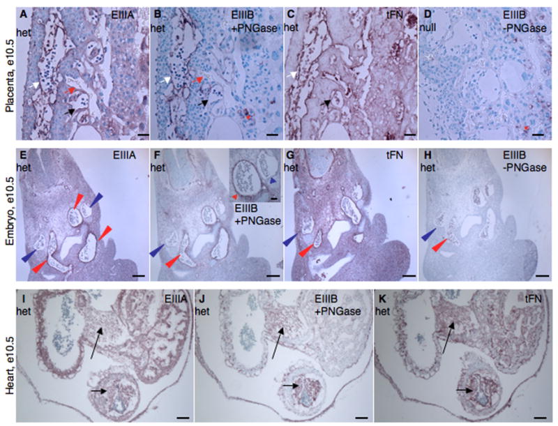
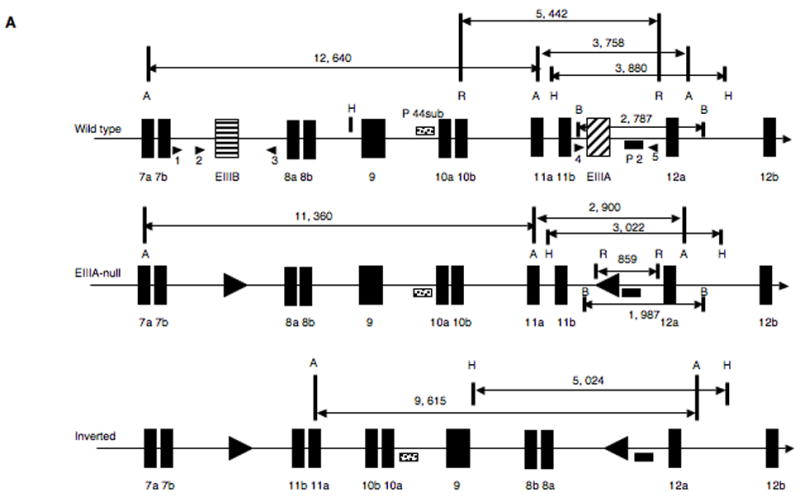
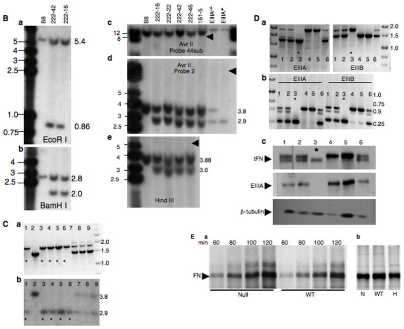
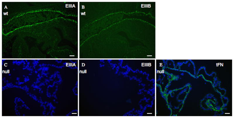
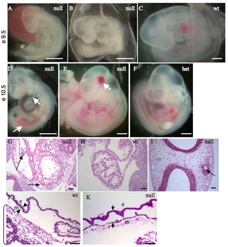
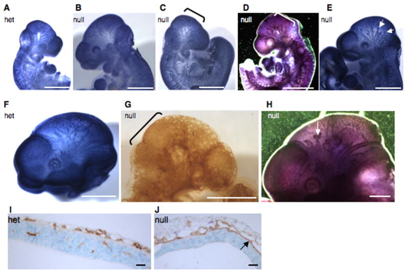
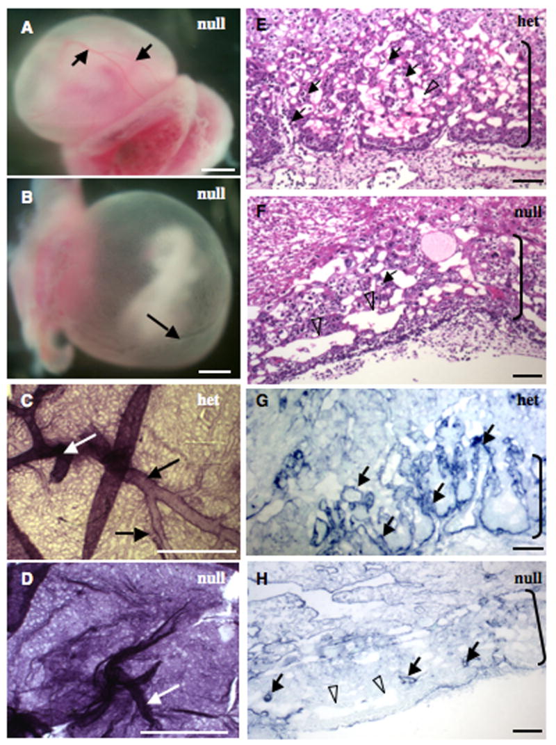
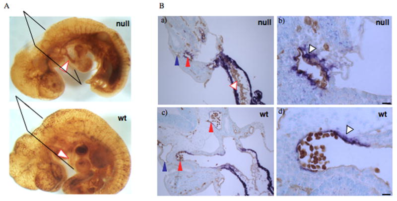
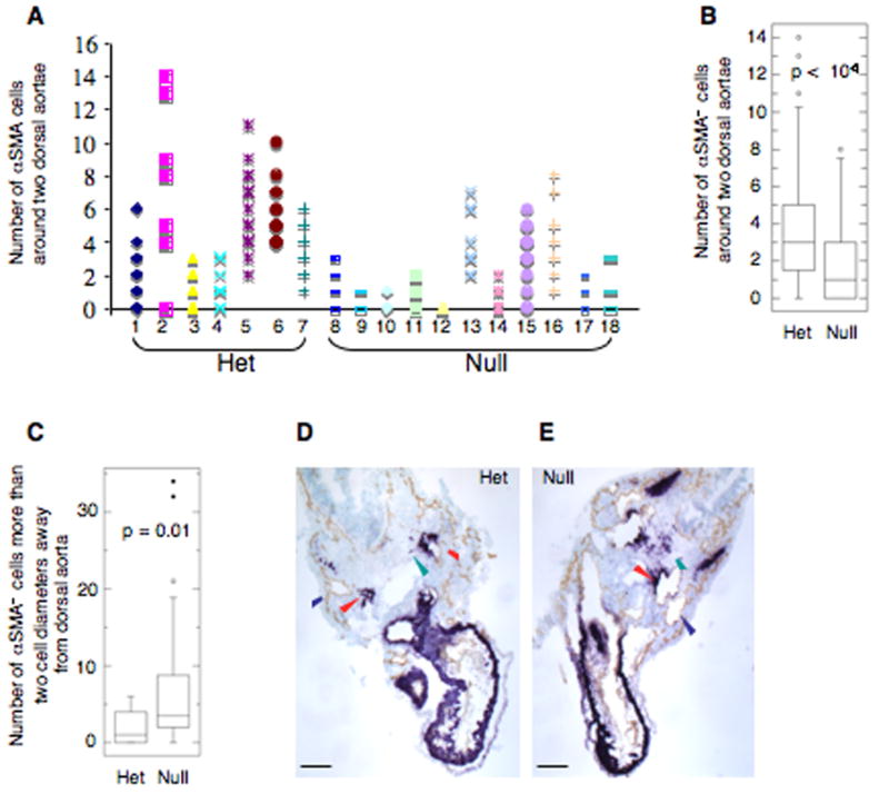
Alternatively spliced variants of fibronectin (FN) containing exons EIIIA and EIIIB are expressed around newly forming vessels in development and disease but are downregulated in mature vasculature. The sequences and patterns of expression of these splice variants are highly conserved among vertebrates, suggestive of their biological importance; however the functions of EIIIA and EIIIB-containing FNs are unknown. To understand the role(s) of these splice variants, we deleted both EIIIA and EIIIB exons from the FN gene and observed embryonic lethality with incomplete penetrance by embryonic day 10.5. Deletion of both EIIIA and EIIIB exons did not affect synthesis or cell surface deposition of FN, indicating that embryonic lethality was due specifically to the absence of EIIIA and EIIIB exons from FN. EIIIA/EIIIB double-null embryos displayed multiple embryonic cardiovascular defects, including vascular hemorrhage, failure of remodeling embryonic and yolk sac vasculature, defective placental angiogenesis and heart defects. In addition, we observed defects in coverage and association with dorsal aortae of alpha-smooth-muscle-actin-positive cells. Our studies indicate that the presence or absence of EIIIA and EIIIB exons alters the function of FN and demonstrate the requirement for these alternatively spliced exons in cardiovascular development.


Figures

Figure 1
Figure 1. Expression of EIIIA and EIIIB splice variants of fibronectin during embryonic development
A–D. Placenta. EIIIA (A), EIIIB (B) and tFN (C) proteins are expressed around embryonic but not maternal blood vessels. D. EIIIA/EIIIB-double-null placenta stained for EIIIB. White arrows point to embryonic blood vessels. Red arrows point to maternal blood vessels. Black arrows point to embryonic vessels lacking FN staining. Red asterisks mark non-specific staining (both EIIIB and PNGase-independent background staining (Peters et al., 1996)). Scale bars are 30 μm. E–H. Embryo. tFN as well as EIIIA and EIIIB splice variants are enriched around arteries but not around veins. Red arrowheads point to arterial vessels and blue arrowheads point to anterior cardinal veins. E. EIIIA, F. EIIIB, G. tFN, H. Control – Staining for EIIIB+-FN without PNGase treatment. Scale bars are 130 μm. I–K. Heart. EIIIA (I), EIIIB (J) and tFN (K) are expressed in cushions (arrows) and endocardium of the heart. Scale bars are 65 μm.
Figure 2
Figure 2. Generation of the EIIIA/EIIIB-double-null allele
A. Generation of the EIIIAEIIIB allele. Top panel represents wild-type locus. A=AvrII, B = BamHI, H=HindIII, R = EcoRI. Arrowheads designate PCR primers used for genotyping. Middle panel represents the correctly targeted EIIIAEIIIB allele. Bottom panel shows the possible inverted allele. B. Southern blots. Marker and band sizes are in kilobases (kb). Blots in all panels except panel c were performed with probe 2; in panel c, probe 44sub was used. a. EcoRI digestion. B8 are EIIIB-null ES cells with the wild-type EIIIA+ allele, lane numbers are names of targeted ES cell clones. The 0.86 kb band corresponds with the EIIIA deletion. b. BamHI digestion. The 1.9 kb band corresponds with the EIIIA deletion. c and d. AvrII digests. The 2.9 kb fragment in d corresponds with the EIIIA deletion. EIIIA−/− and EIIIA+/− cells were used as controls. e. HindIII digest. The 3 kb band corresponds with the EIIIA deletion. Arrowheads in c–e point to where bands corresponding with the possible inverted allele would have been located. C. Genomic PCR for EIIIB allele (a) and a Southern blot (b, AvrII, probe 2) for EIIIA allele from a litter of nine e9.5 embryos derived from EIIIAEIIIB/EIIIA+EIIIB+ intercross. Asterisks denote double-null embryos. D. a. Genomic PCR to detect EIIIA+, EIIIB+, EIIIA, and EIIIB alleles, b. RT-PCR to detect EIIIA+, EIIIB+, EIIIA, and EIIIB mRNA, and c. Western blot to detect tFN, EIIIA and βtubulin from a litter of six e9.5 embryos derived from EIIIAEIIIB/EIIIA+EIIIB+ intercross. Asterisks denote double-null embryos. E. a. Time-course of FN secretion into the medium and b. FN incorporation into the extracellular matrix of metabolically labeled wild-type, double-null and double-het MEFs.
Figure 3
Figure 3. EIIIA/EIIIB-double-null embryos express only EIIIA and EIIIB FN
A. Sagittal sections through e9.5 embryos show expression of EIIIA+ and B. EIIIB+ -FN in wild-type littermate control embryos. The vessel stained is dorsal aorta. C – E. Yolk sacs of e 9.5 EIIIA/EIIIB-double-null embryos show lack of EIIIA (C) and EIIIB (D) expression, whereas FN (E, green) is expressed. DAPI was used to stain nuclei. Scale bars are 30 μm.
Figure 4
Figure 4. Phenotypes of EIIIA/EIIIB-double-null embryos
A–C Representative double-null (A, B) and wild-type (C) embryos at day e9.5 of development are shown. Note hemorrhage, truncated posterior and a kinked neural tube in A, and avascular and blistered yolk sac in B. D–F. Representative double-null (D, E) and wild-type (F) embryos at e10.5 are shown. Note shortened and hemorrhagic posterior and a thinned outflow tract (arrows) in D and a localized head hemorrhage (arrow) in E. Scale bars are 1 mm. G–I. Sagittal sections through embryonic hearts at e9.5 (G, H) and a transverse section through an e10.5 embryonic head (I). Arrows in G point to blood in pericardial cavity as well as in the space between endocardial and muscle layers; arrows in I points to hemorrhage in the head. Scale bars are 30 μm. J, K. Sections through wild-type and double-null yolk sacs. Arrowheads mark mesodermal (m) and endodermal (e) layers. Note the separation of these layers in K. Bracket marks placenta in J.
Figure 5
Figure 5. Head vasculature in double-null embryos at e9.5 and e10.5
A–E. Pecam-1 staining of e9.5 heads. Note a regular pattern of small and large blood vessels in a heterozygous (A) and in a grossly normal double-null embryo (B), compared with a syncytial appearance of small head vessels in a morphologically defective (C) and a grossly normal (D) double-null embryo. Note blunt-ended and interrupted vessels in another grossly normal double-null embryo (E). F–H. Pecam-1 staining of e10.5 heads. Note a regular pattern of small and large vessels in a heterozygous (F) embryo as opposed to syncytial appearance of small vessels and the scarcity of large vessels in a morphologically abnormal double-null embryo (G), as well as short, blunt-ended and interrupted head vessels in a grossly normal double-null (H). Brackets highlight syncytial endothelial sheets in C, D and G and arrows point to blunt-ended vessels in E and H. Scale bars are 1 mm in all except G, where scale bar is 240 μm. I–J. Transverse sections through heads of a heterozygous (I) and a double-null (J) embryo at e10.5 show the presence of small vessels in the head and vessel invasion into the neural fold in the heterozygote (I). In the double-null (J) the vessels appear dilated, resulting from two sheets of endothelial cells touching each other in some places. Also note the lack of vascular invasion into the neural fold in the double-null (J). Scale bars are 65 μm.
Figure 6
Figure 6. Extraembryonic vascular defects in EIIIA/EIIIB-double-null embryos
A–D. Yolk sacs. A, B. EIIIA/EIIIB-double-null yolk sacs showing normal (A) and defective (B) vasculature. Yolk sac in A has large (arrows) and small blood vessels while the one in B has only one large vessel (arrow) and abnormally patterned small vessels. C–D. A heterozygous and a double-null yolk sac stained with antibody to Pecam-1. White arrows point to vitelline vessels and black arrows point to large yolk sac blood vessels. Notice the size and the regular pattern of yolk sac vessels in C, and syncytial appearance of vasculature and absence of large blood vessels in D. Scale bars are 1 mm. E–H. Placental labyrinths (brackets) in heterozygous (E and G) and double-null (F and H) embryos. Note extensive embryonic vasculature permeating the labyrinthine layer in E and the scarcity of embryonic blood vessels in F. Filled arrows point to embryonic and open arrowheads to maternal vessels. G, H. Vascular pattern in the labyrinth layer is revealed with an antibody to laminin1. Note abundant finger-like vascular projections in heterozygous (G) but not double-null (H) placenta. Scale bars are 65 μm.
Figure 7
Figure 7. Defects in heart cushion formation and in association of αSMA cells with dorsal aortae of e10.5 embryos
A. Whole mount Pecam-1 stain revealing blood vessel pattern. Arrowheads point to outflow tracts in double-null and wild-type embryos. Notice a thin outflow tract as well as abundant Pecam-1 reactivity within the outflow tract of the double-null embryo as compared with the wild-type embryo. B. Transverse sections through embryonic hearts. Notice multilayering of endothelial cells in a) compared with c). Scale bars are 130 μm. b and d. αSMA cells are closely associated with dorsal aorta in the wild-type embryos while in the double-null embryo, the αSMA cells are rounded and not well spread around the vessel. Scale bars are 15 μm.
Figure 8
Figure 8. Quantification of αSMA cells around dorsal aortae at e9.5 and e10.5
αSMA cells around each of the two dorsal aortae were enumerated. A. 97 serial sections from seven heterozygous embryos representing four different litters and 126 serial sections from eleven double-null embryos representing five different litters were examined. The numbers of αSMA cells per section around the two dorsal aortae are plotted. Individual embryos are presented along the abscissa. Embryo number 10 was anemic. B. Summary of the results in A presented as a box plot (the box contains the middle 50% of the data, the horizontal line within the box is the median, points outside of the whiskers (10–90% of data) are outliers). C. Delay in recruitment of αSMA cells to dorsal aortae in embryos at e10.5 dpc. Three out of five closely examined, morphologically defective double-null embryos representing three different litters had a defect in recruitment of αSMA+ cells to dorsal aortae. D, E. Transverse sections through a Heterozygous and a double-null embryo stained with antibodies to Pecam-1 and αSMA show excess of αSMA+ cells outside of the immediate proximity of the dorsal aortae. Blue and red arrowheads point to veins and arteries respectively, green arrowhead points to αSMA+ cells. Scale bars are 130 μm.

References

    1. Anson-Cartwright L, Dawson K, Holmyard D, Fisher SJ, Lazzarini RA, Cross JC. The glial cells missing-1 protein is essential for branching morphogenesis in the chorioallantoic placenta. Nat Genet. 2000;25:311–4. - PubMed
    1. Aota S, Nagai T, Yamada KM. Characterization of regions of fibronectin besides the arginine-glycine-aspartic acid sequence required for adhesive function of the cell-binding domain using site-directed mutagenesis. J Biol Chem. 1991;266:15938–43. - PubMed
    1. Astrof S, Crowley D, George EL, Fukuda T, Sekiguchi K, Hanahan D, Hynes RO. Direct test of potential roles of EIIIA and EIIIB alternatively spliced segments of fibronectin in physiological and tumor angiogenesis. Mol Cell Biol. 2004;24:8662–70. - PMC - PubMed
    1. Bloom L, Ingham KC, Hynes RO. Fibronectin regulates assembly of actin filaments and focal contacts in cultured cells via the heparin-binding site in repeat III13. Mol Biol Cell. 1999;10:1521–36. - PMC - PubMed
    1. Borsi L, Balza E, Bestagno M, Castellani P, Carnemolla B, Biro A, Leprini A, Sepulveda J, Burrone O, Neri D, et al. Selective targeting of tumoral vasculature: comparison of different formats of an antibody (L19) to the ED-B domain of fibronectin. Int J Cancer. 2002;102:75–85. - PubMed

Publication types