. 2007 Oct 1;313(16):3658-70.
doi: 10.1016/j.yexcr.2007.07.007. Epub 2007 Jul 19.

The Parkinson's disease-associated protein, leucine-rich repeat kinase 2 (LRRK2), is an authentic GTPase that stimulates kinase activity

Affiliations

The Parkinson's disease-associated protein, leucine-rich repeat kinase 2 (LRRK2), is an authentic GTPase that stimulates kinase activity

Luxuan Guo et al. Exp Cell Res. .

Abstract

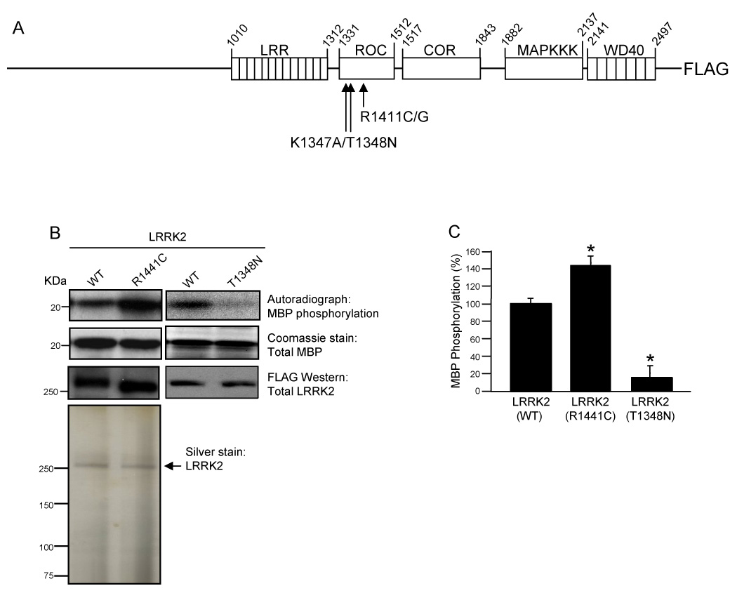
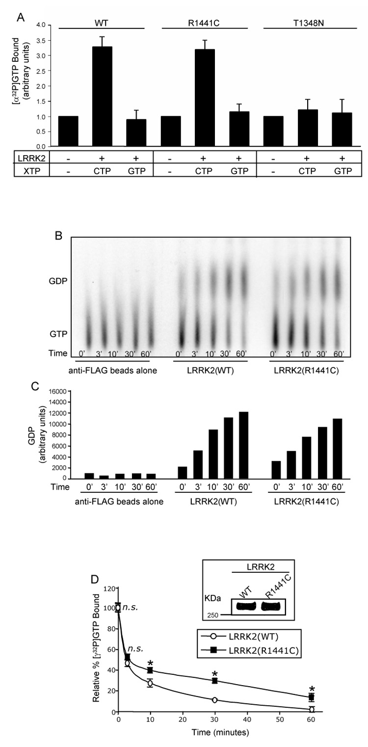
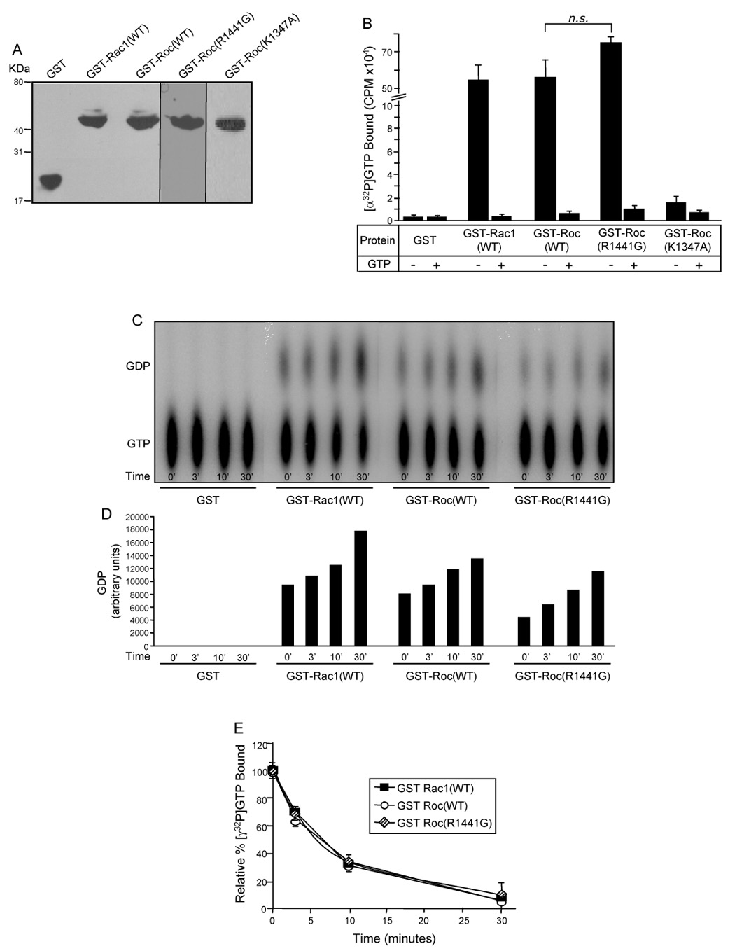
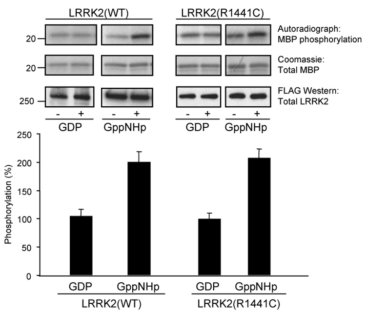
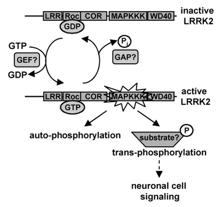
Mutations in the leucine-rich repeat kinase 2 (LRRK2) gene are the leading cause of autosomal dominant Parkinson's disease (PD). LRRK2, a member of the ROCO protein family, contains both Ras GTPase-like (Roc) and kinase (MAPKKK) domains, as well as other functional motifs. Here, we have identified LRRK2 as the first mammalian ROCO protein that is an authentic and functional GTPase, defined by the ability to bind GTP and undergo intrinsic GTP hydrolysis. Furthermore, the Roc domain is sufficient for this native GTPase activity and binds and hydrolyzes GTP indistinguishably from the Ras-related small GTPase, Rac1. The PD-associated mutation, R1441C, located within the Roc domain, leads to an increase in LRRK2 kinase activity and a decrease in the rate of GTP hydrolysis, compared to the wild-type protein, in an in vitro assay. This finding suggests that the R1441C mutation may help stabilize an activated state of LRRK2. Additionally, LRRK2-mediated phosphorylation is stimulated upon binding of non-hydrolyzable GTP analogs, suggesting that LRRK2 is an MAPKKK-activated intramolecularly by its own GTPase. Since GTPases and MAPKKKs are upstream regulators of multiple signal transduction cascades, LRRK2 may play a central role in integrating pathways involved in neuronal cell signaling and the pathogenesis of PD.


Figures

Fig. 1
Fig. 1. The R1441C PD-associated mutation augments LRRK2 kinase activity
(A) Schematic diagram of LRRK2 multi-domain structure. LRRK2 contains several functional domains including N-terminal leucine-rich repeats (LRRs), a Roc (Ras of complex protein) domain, a COR (C-terminal of Roc) domain, a mitogen-activated protein kinase kinase kinase (MAPKKK) domain and C-terminal WD40 repeats. Full-length wild-type and mutant LRRK2 proteins were cloned in frame into a modified pCEP4 vector containing a C-terminal FLAG epitope tag. Arrows indicate the location of the K1347A, T1348N and R1441C/G mutations, within the Roc domain. (B) Phosphorylation of MBP by LRRK2. Autoradiographs show incorporated 32P in MBP resolved by 12% Tris-glycine SDS-PAGE. MBP levels were determined by Coomassie staining. Immunoprecipitated LRRK2 was resolved by Novex 6% SDS-PAGE and LRRK2 protein levels were examined by anti-FLAG Western blot analysis. The purity of LRRK2 immunoprecipitates was analyzed by silver staining. (C) Quantification of phosphorylation of MBP by LRRK2. Autoradiographs of 32P incorporated MBP were quantified by densitometry and normalized to LRRK2 protein levels. Wild-type LRRK2 phosphorylation activity was set to 100%. Error bars for part C represent standard error of the mean (SEM) for three independent experiments. * indicates p<0.01 compared to wild-type LRRK2, assessed by a two-tailed unpaired Student’s t-test.
Fig. 2
Fig. 2. LRRK2 is a functional GTPase and the R1441C PD-associated mutation has a decreased rate of GTP hydrolysis
(A) GTP-binding to LRRK2. LRRK2-FLAG immunoprecipitates were assayed for [α-32P]GTP binding in the presence of competing nucleotides (unlabeled CTP or GTP). CPM of [α-32P]GTP bound were normalized to background levels of the negative control (set to 1, arbitrary units) and plotted. Negative control represents immunoprecipitates of anti-LRRK2 antibody coupled to Dynabeads prepared from untransfected cell lysates. Error bars represent SEM for three independent experiments. (B) GTP hydrolysis of LRRK2 analyzed by the TLC method. Immunoprecipitated wild-type and R1441C mutant LRRK2 were assayed for [α-32P]GTP hydrolysis at 0, 3, 10, 30 and 60mins by spotting on TLC plates. The anti-FLAG beads alone negative control represents immunoprecipitates of anti-FLAG beads prepared from untransfected cell lysates. The autoradiograph shown is representative of five independent experiments. (C) Quantification of GTP hydrolysis of LRRK2 analyzed by the TLC method. Amounts of GDP on the representative autoradiograph, shown in Fig. 2B, were quantified by densitometry. (D) GTP hydrolysis of LRRK2 analyzed by the infinite dilution and rapid filtration method. Inset shows anti-FLAG Western blot of immunoprecipitated LRRK2 proteins to demonstrate equivalent amounts of protein utilized in the GTP hydrolysis assay. Immunoprecipitated wild-type and R1441C mutant LRRK2 were assayed for GTP hydrolysis by incubation with [γ-32P]GTP and equivalent aliquots were removed at 0, 3, 10, 30 and 60mins. The relative percent of [γ-32P]GTP bound, after subtracting background binding to FLAG beads alone (negative control), were plotted. CPM of [γ-32P]GTP bound at time zero was set to 100%. Error bars represent SEM for one experiment performed in triplicate. Similar results were observed in two independent experiments performed in triplicate. * indicates p<0.01 and n.s. is non-significant compared to wild-type LRRK2, assessed by a two-tailed unpaired Student’s t-test.
Fig. 3
Fig. 3. The Roc domain is responsible for LRRK2 GTPase activity
(A) Western blot of GST-fusion proteins. Purified GST fusion proteins were resolved by 12.5% Tris-glycine SDS-PAGE, transferred to PVDF membrane and Western blotted with anti-GST antibody to demonstrate equivalent amounts of protein utilized in the GTP-binding and hydrolysis assays. (B) GTP-binding to the GST-Roc domain of LRRK2. Purified recombinant GST-fusion proteins were assayed for [α-32P]GTP binding in the absence (−) or presence (+) of competing nucleotide (unlabeled GTP). CPM of [α-32P]GTP bound were plotted. (C) GTP hydrolysis of the GST-Roc domain of LRRK2 analyzed by the TLC method. GST-fusion proteins were assayed for [α-32P]GTP hydrolysis at 0, 3, 10 and 30mins by spotting on TLC plates. The autoradiograph shown is representative of three independent experiments. (D) Quantification of GTP hydrolysis of the GST-Roc domain of LRRK2 – TLC method. Amounts of GDP on the representative autoradiograph, shown in Fig. 3C, were quantified by densitometry. (E) GTP hydrolysis of the GST-Roc domain of LRRK2 – infinite dilution and rapid filtration method. GST-fusion proteins were incubated with [γ-32P]GTP and equivalent aliquots were removed at 0, 3, 10 and 30mins. The relative percent of [γ-32P]GTP bound, after subtracting background binding to GST alone (negative control), were plotted. CPM of [γ-32P]GTP bound at time zero was set to 100%. For all experiments GST and GST-Rac1(WT) were used as negative and positive controls, respectively. Error bars represent SEM for one experiment performed in triplicate. Similar results were observed in three independent experiments performed in triplicate. n.s. is non-significant compared to GST-Roc(WT), assessed by a two-tailed unpaired Student’s t-test.
Fig. 4
Fig. 4. LRRK2 kinase activity is stimulated by GTP-binding
Immunoprecipitated LRRK2 was assayed for phosphorylation of MBP in the absence or presence of 5 µM of either GDP or a non-hydrolyzable GTP analog, Gpp(NH)p. Autoradiographs demonstrate incorporated 32P in MBP resolved by 12% Tris-glycine SDS-PAGE (top panel). MBP levels were determined by Coomassie staining (middle panel) and LRRK2 protein levels were determined by Western blot analysis using anti-FLAG antibody (bottom panel). Autoradiographs of 32P incorporated MBP (top panel) were quantified by densitometry and normalized to LRRK2 protein expression levels (bottom panel). LRRK2 trans-phosphorylation activity in the presence of GDP was set to 100%. Error bars represent SEM for three independent experiments.
Fig. 5
Fig. 5. Proposed model for the GTP-dependent activation of LRRK2 kinase activity
LRRK2 is likely to cycle between an inactive (GDP-bound) and active (GTP-bound) conformation within the cell. GTP-binding to the Roc domain likely activates LRRK2 by stimulating kinase activity. This exchange of GDP for GTP may potentially be facilitated by a putative GEF. In the active conformation, LRRK2 is poised to undergo auto-phosphorylation and potentially phosphorylate other cellular substrates to initiate neuronal cell signaling cascades. Once GTP is hydrolyzed to GDP, potentially aided by a putative GAP, LRRK2 returns to its inactive state in which kinase activity is terminated.

References

    1. Tanner CM, B-S Y. Epidemiology of Parkinson's disease. Adv Neurol. 1999;80:153–159. - PubMed
    1. Moore DJ, W A, Dawson VL, Dawson TM. Molecular pathophysiology of Parkinson's disease. Annu Rev Neurosci. 2005;28:57–87. - PubMed
    1. Cookson MR. The biochemistry of Parkinson's disease. Annu Rev Biochem. 2005;74:29–52. - PubMed
    1. Paisan-Ruiz C, J S, Evans EW, Gilks WP, Simon J, van der Brug M, Lopez de Munain A, Aparicio S, Gil AM, Khan N, Johnson J, Martinez JR, Nicholl D, Carrera IM, Pena AS, de Silva R, Lees A, Marti-Masso JF, Perez-Tur J, Wood NW, Singleton AB. Cloning of the gene containing mutations that cause PARK8-linked Parkinson's disease. Neuron. 2004;44:595–600. - PubMed
    1. Zimprich A, B S, Leitner P, Lichtner P, Farrer M, Lincoln S, Kachergus J, Hulihan M, Uitti RJ, Calne DB, Stoessl AJ, Pfeiffer RF, Patenge N, Carbajal IC, Vieregge P, Asmus F, Muller-Myhsok B, Dickson DW, Meitinger T, Strom TM, Wszolek ZK, Gasser T. Mutations in LRRK2 cause autosomal-dominant parkinsonism with pleomorphic pathology. Neuron. 2004;44:601–607. - PubMed

Publication types

MeSH terms

Substances