. 2007 Aug 17;27(4):573-84.
doi: 10.1016/j.molcel.2007.06.022.

Activator-mediated recruitment of the MLL2 methyltransferase complex to the beta-globin locus

Affiliations

Activator-mediated recruitment of the MLL2 methyltransferase complex to the beta-globin locus

Celina Demers et al. Mol Cell. .

Abstract

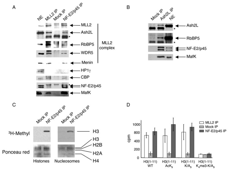
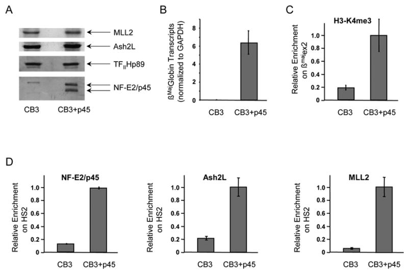
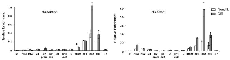
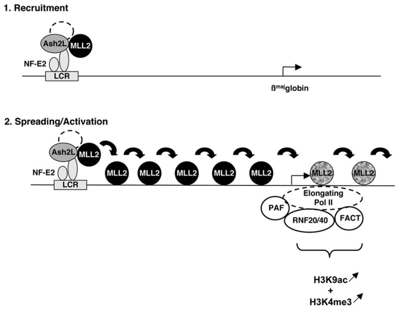
MLL-containing complexes methylate histone H3 at lysine 4 (H3K4) and have been implicated in the regulation of transcription. However, it is unclear how MLL complexes are targeted to specific gene loci. Here, we show that the MLL2 complex associates with the hematopoietic activator NF-E2 in erythroid cells and is important for H3K4 trimethylation and maximal levels of transcription at the beta-globin locus. Furthermore, recruitment of the MLL2 complex to the beta-globin locus is dependent upon NF-E2 and coincides spatio-temporally with NF-E2 binding during erythroid differentiation. Thus, a DNA-bound activator is important initially for guiding MLL2 to a particular genomic location. Interestingly, while the MLL2-associated subunit ASH2L is restricted to the beta-globin locus control region 38 kb upstream of the beta(maj)-globin gene, the MLL2 protein spreads across the beta-globin locus, suggesting a previously undefined mechanism by which an activator influences transcription and H3K4 trimethylation at a distance.


Figures

Figure 1
Figure 1. NF-E2 associates with the H3K4 methyltransferase complex MLL2 in differentiated erythroid nuclear extracts
(A) Western blot analysis of proteins immunoprecipitated from an erythroid nuclear extract using Abs against MLL2 and NF-E2/p45. A mock IP using normal rabbit IgG was used as a negative control. Abs used for western blot are indicated on the right. *, Ab heavy chain (B) Western blot analysis of proteins immunoprecipitated from an erythroid nuclear extract using Abs against Ash2L. *, Ab heavy chain (C) NF-E2/p45 and Mock immunoprecipitated proteins were used in an in vitro methylation assay in the presence of [3H] SAM and equal amounts of purified core histones (left) or nucleosomes (right) as a substrate. The top panel is an autoradiograph of the bottom panel (stained with Ponceau red). (D) Eleven amino-acids peptides corresponding to the N-term part of histone H3 that are either wild-type (WT), acetylated at lysine 9 (AcK9), mutated at position 9 (Mut. K/A9) or mutated at lysine 9 and trimethylated at lysine 4 (K4me3-K/A9) were used as a substrate for methylation by NF-E2/p45-, mock-, or MLL2- associated proteins and the radioactivity was counted (in cpm) after a filter binding assay. Error bars correspond to the standard deviation of 3 counts of 1 min. each. The experiment was performed at least twice independently.
Figure 2
Figure 2. Recruitment of the MLL2 complex and H3K4 trimethylation at the β-globin locus are dependent upon NF-E2/p45
(A) Western blot analysis of nuclear extracts prepared from the NF-E2/p45-null mouse erythroid cell line CB3 and from a rescued clone derived from this cell line, which expresses exogenous NF-E2/p45 (CB3+p45). CB3 cells and the rescued clone were induced to differentiate along the erythroid lineage by exposure to DMSO. (B) NF-E2/p45 expression in the CB3 cell line partially restores β-globin transcription. Production of βmaj-globin mRNA transcripts after erythroid differentiation was measured by RT-qPCR in the NF-E2/p45 null CB3 cell line and in a rescued NF-E2/p45-expressing CB3 clone. βmaj-globin transcripts were normalized for the amount of GAPDH transcripts. Average values of RT-qPCR triplicates are indicated with error bars corresponding to standard deviations. (C) H3K4me3 enrichment at the βmaj-globin gene was measured by chromatin immunoprecipitation (ChIP) in the absence (CB3) vs presence (CB3+p45) of NF-E2/p45 (D) NF-E2/p45, Ash2L and MLL2 binding to the β-globin locus were analyzed by ChIP in the absence (CB3) vs presence (CB3+p45) of NF-E2/p45. TaqMan probes used to reveal the ChIPs by real-time qPCR are the same as in Figure 3. Average values of qPCR duplicates are expressed as a function of the highest enrichment on the locus with error bars corresponding to standard deviations. Each experiment was performed at least twice independently.
Figure 3
Figure 3. Recruitment of NF-E2/p45 and the MLL2 complex to the β-globin locus during erythroid differentiation analyzed by chromatin immunoprecipitation
(A) Schematic representation of the murine β-globin locus. Shaded triangles represents the β-like globin genes. The white triangle represents the inactive olfactory receptor gene. Vertical arrows represent DNAse I hypersensitive sites (HSs). The βmaj-globin and Ey genes are shown in details below the locus with their 3 exons (ex). Positions of the TaqMan probes used to reveal the chromatin immunoprecipitations (ChIPs) by real-time qPCR are indicated relative to the βmaj-globin transcription start site (set to +1). Myog. indicates the muscle-specific gene myogenin. (B) ChIP analysis of NF-E2/p45, Ash2L and MLL2 binding to the β-globin locus during erythroid differentiation. ChIPs were performed in MEL cells before (Nondiff.) and after (Diff.) induction of erythroid differentiation. Average values of qPCR duplicates are expressed as a function of the highest enrichment on the locus with error bars corresponding to standard deviations. Each ChIP experiment was performed at least twice independently.
Figure 4
Figure 4. Enrichment of H3K4me3 and H3K9ac is limited to the 5′ region of the βmaj-globin gene coding area
(A) Analysis of H3K4me3 at the β-globin locus during erythroid differentiation by ChIP. (B) Analysis of H3K9ac at the β-globin locus during erythroid differentiation by ChIP. TaqMan probes used to reveal the ChIP by real-time qPCR are the same as in Figure 3. ChIPs were performed in MEL cells before (Nondiff.) and after (Diff.) induction of erythroid differentiation. Average values of qPCR duplicates are expressed as a function of the highest enrichment on the locus with error bars corresponding to standard deviations. Each ChIP experiment was performed at least twice independently.
Figure 5
Figure 5. Ash2L and MLL2 are required for maximal levels of βmaj-globin transcription and H3K4 trimethylation during erythroid differentiation
(A) Western blot analysis of the doxycyclin (Dox)-inducible knockdowns (KnDs) of Ash2L and MLL2 in differentiated MEL cells. Nuclear extracts were prepared and revealed using the Abs indicated on the right. SAP30 and TFIIHp89 are used as loading controls. (B) Ash2L and MLL2 KnDs lead to a defect in transcription at the β-maj globin gene. Transcription at the βmaj-globin gene was assessed by real-time qRT-PCR before (Nondiff) and after (Diff) erythroid differentiation in Dox-induced (shaded bar) vs non-induced (white bar) MEL cells. The parent clone served as a negative control. βmaj-globin transcription relative to GAPDH is expressed as a percentage of the highest level of transcripts. Average values of RT-qPCR triplicates are indicated with error bars corresponding to standard deviations. Each experiment was performed at least 3 times independently. (C) Ash2L and MLL2 KnDs lead to a defect in H3K4 trimethylation at the β-maj globin gene. H3K4me3 enrichment at the βmaj-globin gene was measured by chromatin immunoprecipitation (ChIP) after erythroid differentiation in Dox-induced (white bar) vs non-induced (shaded bar) MEL cells. TaqMan probes used to reveal the ChIPs by real-time qPCR are the same as in Figure 3. Average values of qPCR duplicates are expressed as a function of the highest enrichment on the locus with error bars corresponding to standard deviations. Each experiment was performed at least twice independently.
Figure 6
Figure 6. Model of recruitment, spreading and activation of the MLL2 methyltransferase complex on the β-globin locus during erythroid differentiation
The MLL2 complex is initially guided to the β-globin Locus Control Region (LCR) via its interaction with the LCR-bound activator NF-E2. Binding of this H3K4 methyltransferase to the LCR does not however result in trimethylation of H3K4 in this region (presumably due to the histone code particular to this region of the locus). Then MLL2 detaches from its core components and spreads across 38 kb where it reaches the elongating Pol II and associated complexes. Interaction with basal transcriptional elongation machinery and/or the presence of specific histone modifications (H3K9ac and/or Ubiquitinated H2B) allows MLL2 to methylate H3K4. The inactive/active MLL2 protein is represented by a black/grey shading. H3K4me3 and H3K9ac are represented below the region where they are enriched on the β-globin locus and the direction of their variation during differentiation is figured by arrows.

Similar articles

Cited by

References

    1. Andrews NC. The NF-E2 transcription factor. Int J Biochem Cell Biol. 1998;30:429–432. - PubMed
    1. Ayton PM, Chen EH, Cleary ML. Binding to nonmethylated CpG DNA is essential for target recognition, transactivation, and myeloid transformation by an MLL oncoprotein. Mol Cell Biol. 2004;24:10470–10478. - PMC - PubMed
    1. Bernstein BE, Kamal M, Lindblad-Toh K, Bekiranov S, Bailey DK, Huebert DJ, McMahon S, Karlsson EK, Kulbokas EJ, 3rd, Gingeras TR, et al. 2005 - PubMed
    1. Genomic maps and comparative analysis of histone modifications in human and mouse. Cell . 120:169–181. - PubMed
    1. Brand M, Ranish JA, Kummer NT, Hamilton J, Igarashi K, Francastel C, Chi TH, Crabtree GR, Aebersold R, Groudine M. Dynamic changes in transcription factor complexes during erythroid differentiation revealed by quantitative proteomics. Nat Struct Mol Biol. 2004;11:73–80. - PubMed

Publication types

MeSH terms

LinkOut - more resources