. 2007 Aug 17;27(4):609-21.
doi: 10.1016/j.molcel.2007.07.024.

A histone H2A deubiquitinase complex coordinating histone acetylation and H1 dissociation in transcriptional regulation

Affiliations

A histone H2A deubiquitinase complex coordinating histone acetylation and H1 dissociation in transcriptional regulation

Ping Zhu et al. Mol Cell. .

Abstract

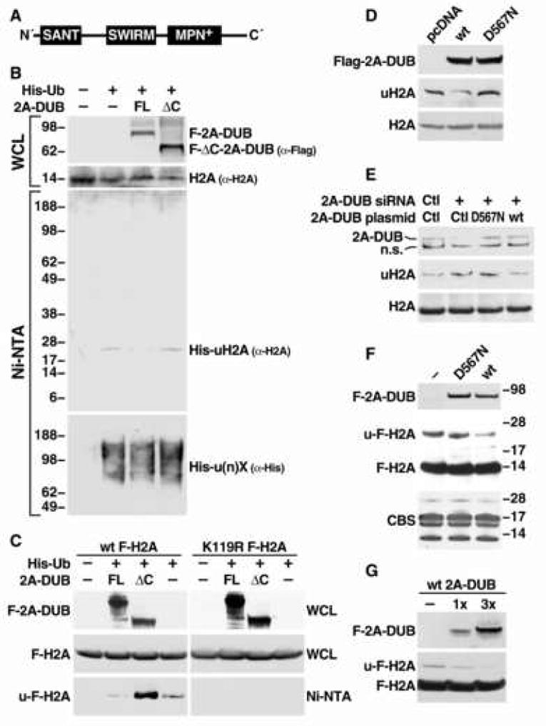
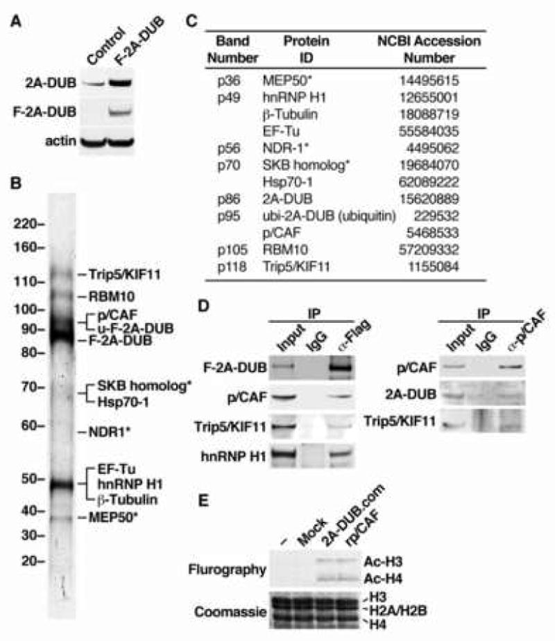
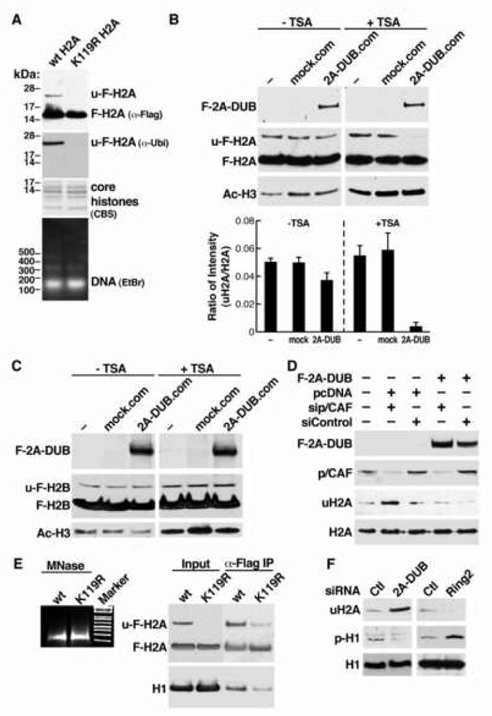
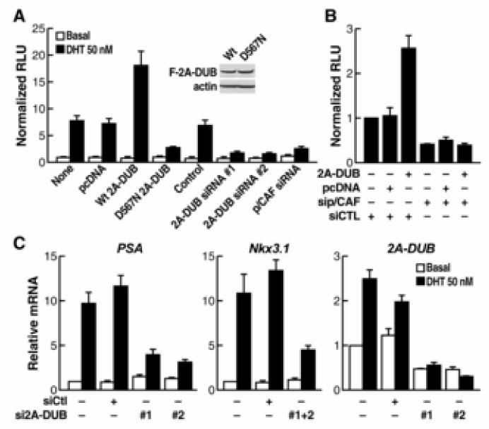
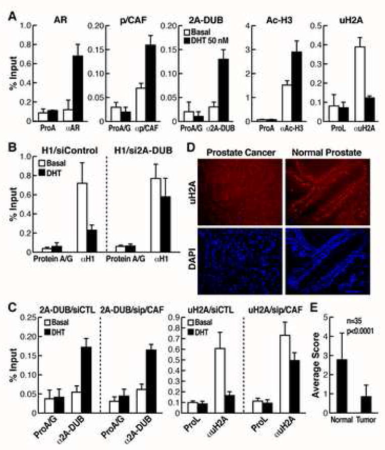
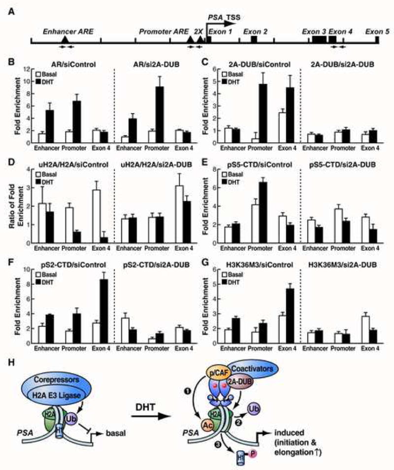
Deciphering the epigenetic "code" remains a central issue in transcriptional regulation. Here, we report the identification of a JAMM/MPN(+) domain-containing histone H2A deubiquitinase (2A-DUB, or KIAA1915/MYSM1) specific for monoubiquitinated H2A (uH2A) that has permitted delineation of a strategy for specific regulatory pathways of gene activation. 2A-DUB regulates transcription by coordinating histone acetylation and deubiquitination, and destabilizing the association of linker histone H1 with nucleosomes. 2A-DUB interacts with p/CAF in a coregulatory protein complex, with its deubiquitinase activity modulated by the status of acetylation of nucleosomal histones. Consistent with this mechanistic role, 2A-DUB participates in transcriptional regulation events in androgen receptor-dependent gene activation, and the levels of uH2A are dramatically decreased in prostate tumors, serving as a cancer-related mark. We suggest that H2A ubiquitination represents a widely used mechanism for many regulatory transcriptional programs and predict that various H2A ubiquitin ligases/deubiquitinases will be identified for specific cohorts of regulated transcription units.


Figures

Figure 1
Figure 1. Identification of a deubiquitinase of histone H2A
(A) Schematic representation of the domain organization of 2A-DUB/KIAA1915/MYSM1 protein. (B) In vivo ubiquitination assay for H2A after overexpression of human full-length (FL) and C-terminal enzymatic JAMM/MPN+ domain-deleted mutant (ΔC) 2A-DUB (aa 17-531) in HEK293T cells. Molecular weight (kDa) is shown on the left. WCL represents whole cell lysates. (C) In vivo ubiquitination assay in HEK293 cells stably expressing wild-type (wt) and K119R Flag-tagged H2A. Anti-Flag antibody was used to detect signals from Flag-H2A. (D) Detection of endogenous uH2A and H2A levels by Western blot analysis after overexpression of Flag-tagged full-length wt and D567N mutant 2A-DUB in HEK293T cells. (E) Detection of endogenous uH2A and H2A levels by Western blotting upon siRNA-mediated knock-down of endogenous 2A-DUB or co-expression of wt or D567N 2A-DUB with siRNA against 2A-DUB. (F) In vitro deubiquitination assay for H2A. Affinity-purified wt and D567N 2A-DUB (transfected into HEK293T cells) were used as enzymatic source (about 2 μg), and acid-extracted histones from wt Flag-H2A-expressing HEK293 cells was used as the substrate (about 1.5 μg). Western analysis of 2A-DUB and Flag-H2A showed the signals of enzymes and substrates, and the Commassie Blue staining (CBS) displayed the loading of core histone substrates. (G) Using 1 μg (1×) or 3 μg (3×) of 2A-DUB in the same assays as (F).
Figure 2
Figure 2. Affinity purification of the 2A-DUB protein complex
(A) Western blot analysis of HEK293 cell lines used for affinity purification. Control and F-2A-DUB cells were stably transfected with an empty vector and a vector encoding Flag-tagged human 2A-DUB, respectively. (B) Silver staining of co-purified peptides in the affinity-purified 2A-DUB protein complex from HEK293 cells after separation by SDS-PAGE. The molecular weight was shown on the left and the identity of the peptide band, analyzed by mass spectrometry, was shown on the right. Asterisks represent bands that were also observed in the materials from the parallel affinity purification of the control cell line, or known to interact with M2 anti-Flag resin. (C) Detailed information of mass spectrometric analysis of the co-purified peptides in the 2A-DUB protein complex. (D) Left panel: co-immunoprecipitation (Co-IP) of 2A-DUB and proteins co-purified with 2A-DUB. Flag-2A-DUB was transiently transfected into HEK293T cells, and immunoprecipitated from the nuclear extracts. Co-immunoprecipitated proteins were detected by Western blot analysis. Right panel: IP using anti-p/CAF to test the co-immunoprecipitated endogenous proteins in HEK293T cells. Inputs: 2%. (E) In vitro histone acetyltransferase (HAT) assay for free core histones using affinity-purified 2A-DUB complex (2A-DUB.com), recombinant HAT domain of p/CAF (rp/CAF) and materials from affinity purification of control cells (mock).
Figure 3
Figure 3. Coordination of histone acetylation, H2A deubiquitination and H1 phosphorylation/dissociation in nucleosomes
(A) Characterization of Flag-H2A-containing mono/oligonucleosomes. Purified nucleosomes from HEK293T cells stably expressing wt and K119R Flag-tagged H2A were evaluated for specificity of the 24-kD ubiquitinated Flag-H2A band (u-F-H2A) by Western blot using anti-Flag and anti-ubiquitin antibodies. Composition of core histones was checked by Coomassie blue staining (CBS), and DNA size of MNase-digested nucleosomes was detected by ethidium bromide (EtBr) staining after gel separation. (B) In vitro deubiquitination assay using affinity-purified 2A-DUB protein complex (2A-DUB.com) and mock purified materials from control cells (mock.com) as the enzymatic source, and using Flag-tagged H2A-containing mono/oligonucleosomes purified from TSA-treated or untreated cells as the substrate. Levels of 2A-DUB, u-F-H2A, F-H2A and acetylation of histone H3 (Ac-H3) were detected by Western blot analysis. Lower panel: quantification of three independent assays. (C) Similar assays as (B), using Flag-tagged H2B-containing mono/oligonucleosomes as the substrate. (D) Flag-2A-DUB and siRNA against p/CAF were transfected into HEK293T cells as indicated. Levels of corresponding proteins were detected by Western blots. (E) Detection of composition of associated linker histone H1 in immuno-purified wt or K119R Flag-H2A-containing mononucleosomes. Mononucleosomal DNA was stained by EtBr (left). (F) Evaluation of phosphorylation of H1 after manipulating uH2A levels by knocking down 2A-DUB or H2A E3 ligase Ring2 in HEK293T cells.
Figure 4
Figure 4. 2A-DUB in activation of androgen/AR-dependent transcription
(A) Reporter assays using an ARE-dependent luciferase reporter in LNCaP prostate cancer cells. Cells were transfected with reporter and different plasmids or siRNA as indicated and then treated with or without AR agonist DHT. The activity of specific luciferase was normalized with that of a co-transfected unregulated renilla luciferase reporter. Values are the relative values to normalized activity from basal, reporter-only transfected cells (bar 1, set as 1), and represent means±SEM of three independent experiments. The similar expression levels of wt and D567N 2A-DUB are shown by Western blot analysis. (B) Assays of ARE-driven luciferase reporter in LNCaP cells. Cells were transfected with indicated plasmids or siRNA in combination with reporters, and then treated with DHT. Normalized relative values are calculated as described in (A). (C) Expression of endogenous AR target genes and 2A-DUB analyzed by quantitative RT-PCR. LNCaP cells were transfected with indicated siRNA, and then treated with DHT if needed. Values (normalized to corresponding values of internal control gene HPRT) are means±SEM of three independent experiments.
Fig. 5
Fig. 5. The role of 2A-DUB and uH2A in androgen signaling in prostate cancer cells
(A) ChIP assays of the promoter of PSA, a target gene of AR. LNCaP cells were cultured in the charcoal-stripped medium and then challenged with DHT for 1 hr. Soluble chromatin was immunoprecipitated by the antibodies indicated, and the bound DNA was analyzed by quantitative PCR using a primer pair flanking the AREs of the PSA promoter. Values (ratios of ChIP to corresponding inputs) are means±SEM of at least two independent experiments. (B) The occupancy of linker histone H1 was detected by quantitative ChIP assays on the promoter of PSA in LNCaP cells, transfected with control or 2A-DUB siRNA. (C) The occupancy of 2A-DUB and uH2A was detected by quantitative ChIP assays on the promoter of PSA in LNCaP cells, transfected with control or p/CAF siRNA. (D) Immunofluorescent staining of uH2A for a tissue microarray of prostate cancer. DAPI staining was performed to visualize the nuclear DNA. One representative staining is shown. (E) Semi-quantification of the uH2A staining by a four-grade scoring system (see Experimental Procedure). Values are mean±SD of grades scored for benign prostate tissues or corresponding prostate tumors. Paired Student's t test was used to calculate the p value.
Figure 6
Figure 6. 2A-DUB in regulation of transcriptional initiation and elongation
(A) Schematic representation of the genomic locus (10 kb) of the PSA gene. Arrows represent the primer sets used for ChIP analysis of the enhancer, promoter and exon 4 region of PSA. TSS: transcription start site. Quantitative ChIP assays were performed for AR (B), 2A-DUB (C), uH2A and H2A (D), PolII (phospho-serine 5 of CTD, pS5-CTD) (E), PolII (phospho-serine 2 of CTD, pS2-CTD) (F) and H3K36M3 (G). LNCaP cells were cultured in the charcoal-stripped medium. Following transfection of control (left panels) or 2A-DUB (right panels) siRNA, cells were treated with DHT for 1 hr, then ChIP was performed as described in Experimental Procedures. Enrichment folds compared to the corresponding control (bead alone) ChIP were calculated and shown. For (D), the ratios of enrichment folds of uH2A to H2A were shown. Values are mean±SD of three biological repeats. One representative of at least two independent experiments is shown. (H) A proposed model of the role of 2A-DUB and uH2A in transcriptional regulation. See the text for details.

Similar articles

Cited by

References

    1. Ambroggio XI, Rees DC, Deshaies RJ. JAMM: a metalloprotease-like zinc site in the proteasome and signalosome. PLoS Biol. 2004;2(1):E2. - PMC - PubMed
    1. Baarends WM, Hoogerbrugge JW, Roest HP, Ooms M, Vreeburg J, Hoeijmakers JH, Grootegoed JA. Histone ubiquitination and chromatin remodeling in mouse spermatogenesis. Dev Biol. 1999;207:322–333. - PubMed
    1. Bannister AJ, Schneider R, Myers FA, Thorne AW, Crane-Robinson C, Kouzarides T. Spatial distribution of di- and tri-methyl lysine 36 of histone H3 at active genes. J Biol Chem. 2005;280(18):17732–17736. - PubMed
    1. Belotserkovskaya R, Oh S, Bondarenko VA, Orphanides G, Studitsky VM, Reinberg D. FACT facilitates transcription-dependent nucleosome alteration. Science. 2003;301(5636):1090–1093. - PubMed
    1. Boyer LA, Latek RR, Peterson CL. The SANT domain: a unique histone-tail-binding module? Nat Rev Mol Cell Biol. 2004;5(2):158–163. - PubMed

Publication types

MeSH terms