. 2007 Aug 17;27(4):636-46.
doi: 10.1016/j.molcel.2007.07.012.

Modulation of p53 function by SET8-mediated methylation at lysine 382

Affiliations

Modulation of p53 function by SET8-mediated methylation at lysine 382

Xiaobing Shi et al. Mol Cell. .

Abstract

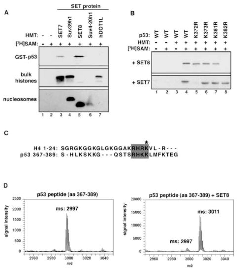
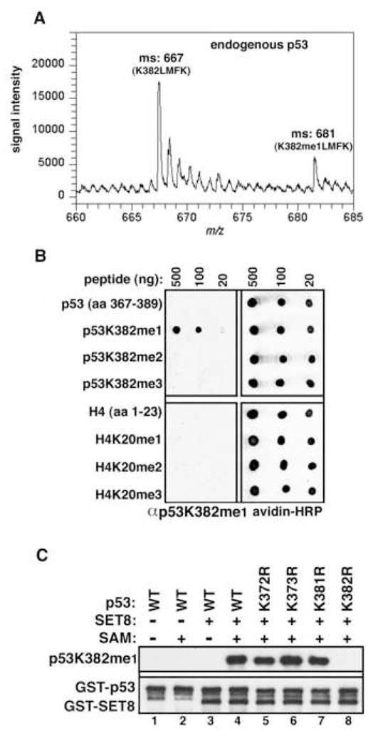
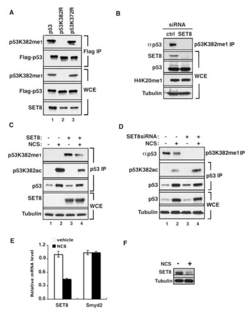
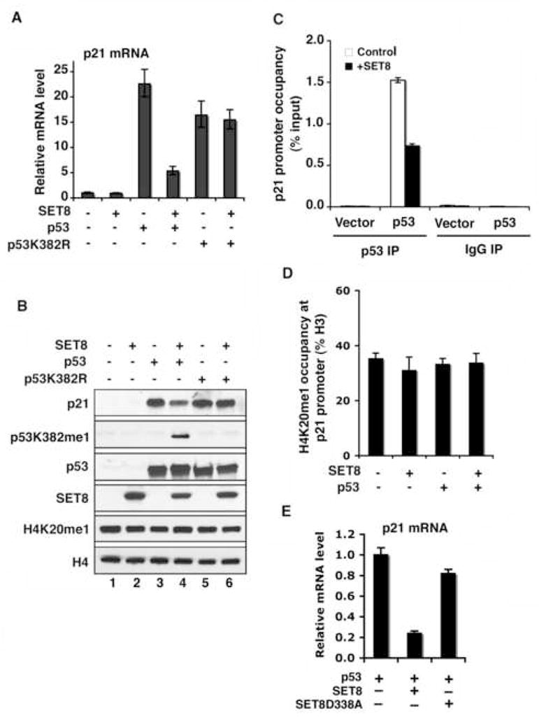
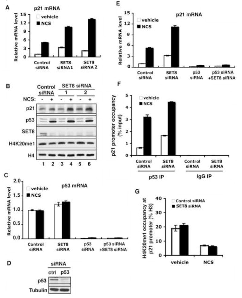
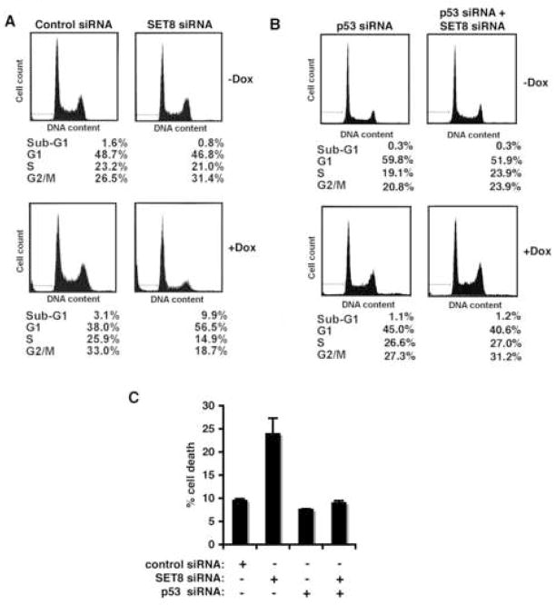
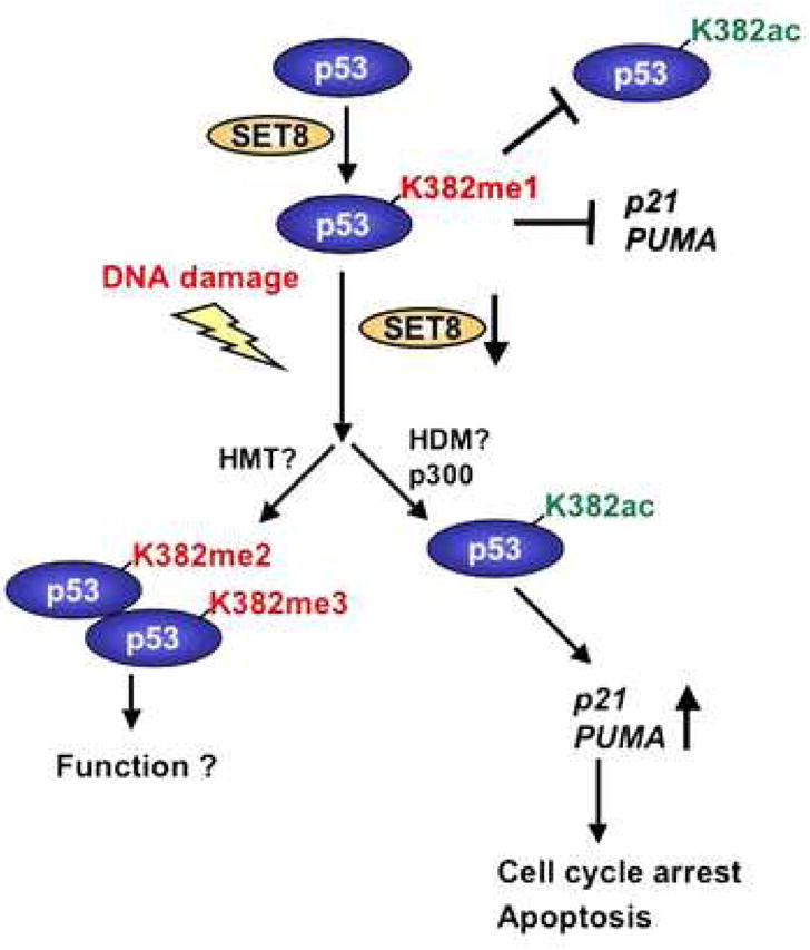
Reversible covalent methylation of lysine residues on histone proteins constitutes a principal molecular mechanism that links chromatin states to diverse biological outcomes. Recently, lysine methylation has been observed on nonhistone proteins, suggesting broad cellular roles for the enzymes generating and removing methyl moieties. Here we report that the lysine methyltransferase enzyme SET8/PR-Set7 regulates the tumor suppressor protein p53. We find that SET8 specifically monomethylates p53 at lysine 382 (p53K382me1). This methylation event robustly suppresses p53-mediated transcription activation of highly responsive target genes but has little influence on weak targets. Further, depletion of SET8 augments the proapoptotic and checkpoint activation functions of p53, and accordingly, SET8 expression is downregulated upon DNA damage. Together, our study identifies SET8 as a p53-modifying enzyme, identifies p53K382me1 as a regulatory posttranslational modification of p53, and begins to dissect how methylation may contribute to a dynamic posttranslational code that modulates distinct p53 functions.


Figures

Figure 1
Figure 1. SET8 monomethylates p53 at K382 in vitro
(A) Identification of SET8 as a novel p53 methyltransferase. Autoradiograms of methyltransferase assays with the indicated recombinant HMTs and substrates. (B) SET8 methylates p53 at K382. Autoradiograms of methyltransferase assays with SET8 or SET7 on wild-type p53 or the indicated p53 mutants. (C) Alignment of amino acid sequences of SET8 substrates histone H4 (aa 1–24) and p53 (aa 367–389). Asterisk indicates SET8 methylation sites on H4 and p53. (D) p53 is monomethylated by SET8 at K382. Mass spectrometry analysis of p53 peptide (aa 367–389) before (left panel) and after (right panel) SET8 methyltransferase reaction.
Figure 2
Figure 2. Characterization of a p53K382me1 modificaiton-specific antibody
(A) Identification of endogenous p53 monomethylated at K382. Mass spectrometry analysis of endogenous p53 IPed from HeLa NE reveals trypsin-digested peptides containing lysine382 (382-KLMFK-386) present in peaks containing either unmethylated or monomethylated K382. (B) Specific recognition of p53K382me1 by the αp53K382me1 antibody. Dot blot analysis of the indicated biotinylated peptides (p53: top; H4: bottom) with αp53K382me1 antibody. Blots were probed with HRP-conjugated streptavidin to control for loading. (C) αp53K382me1 antibody recognizes p53 in vitro methylated at K382 by SET8. Immunoblot analysis of the indicated recombinant p53 protein or mutants ± methyltransferase assays with SET8 as indicated. Total p53 and SET8 were detected with GST antibody to show equal loading.
Figure 3
Figure 3. p53 is monomethylated at K382 in vivo by SET8
(A) Ectopic SET8 specifically methylates p53 at K382 in vivo. Western blot analysis with αp53K382me1 antibody of Flag IPs or WCE (whole cell extracts) from U2OS cells expressing SET8 and the indicated Flag-tagged p53 derivatives. p53 and SET8 protein levels in WCE are shown. (B) Knockdown of endogenous SET8 decreases endogenous levels of p53K382me1 in U2OS cells. Western analysis of p53 levels present in αp53K382me1 IPs of U2OS cells treated with control or SET8 siRNAs. Total p53, SET8, H4K20me1 and tubulin present in the WCE are shown. (C) SET8 negatively regulates acetylation of p53 at K382. Western analysis with αp53K382me1, p53K382ac and p53 antibodies of p53 IPs from U2OS cells transfected with control vector or SET8, and treated for 2 hrs with 0.5 μg/ml NCS. SET8 and tubulin levels in the WCE are shown. Endogenous p53K382me1 is observed with longer exposure (Supplementary Figure 2C) (D) p53K382me1 levels decrease upon DNA damage. Western analyses with the indicated antibodies of αp53K382me1 and p53 (DO1) IPs from U2OS cells transfected with control or SET8 siRNA and treated with NCS for 2 hrs. Tubulin and total p53 levels present in WCE are shown to control for loading. (E) SET8 mRNA expression decreases in response to DNA damage. Real-time PCR analysis of SET8 and Smyd2 mRNA levels present in U2OS cells ± NCS treatment (0.5 μg/ml, 4 hrs). (F) SET8 protein levels decrease in response to DNA damage. Western analysis of SET8 in U2OS cells as in (E).
Figure 4
Figure 4. SET8 methylation of p53 at K382 suppresses p53 transactivation activity
(A) and (B) SET8 inhibits induction of p21 transcription (A) and p21 protein expression (B) by wild-type p53, but does not affect the activity of the p53K382R mutant. (A) Real-time PCR analyses of p21 mRNA levels in H1299 cells transfected with control vector, p53 or p53K382R mutant, ± SET8. (B) Western analyses with the indicated antibodies of H1299 cells WCE as in (A). (C) SET8 expression attenuates occupancy of p53 at the p21 promoter. p53 occupancy at the p21 promoter in H1299 cells transfected with control vector or p53, ± SET8 was determined by ChIP analyses. DO-1 antibody was used for p53 ChIP and IgG was used as control. Occupancy values (ChIP/inputX100) were determined by real-time PCR. (D) SET8 expression does not alter H4K20me1 levels at the p21 promoter. Occupancy of H4K20me1 (H4K20me1 ChIP/H3 ChIPX100) at the p21 promoter was determined as in (C). Error bars in (A, C, D) indicate ± s.e.m. from three experiments. (E) SET8 catalytic mutant SETD338A fails to suppress p53 transactivation activity on target genes. Real-time PCR analyses of relative p21 mRNA levels in H1299 cells co-transfected with p53, SET8, SET8D338A or control vector as indicated.
Figure 5
Figure 5. SET8 RNAi augments p53 activity in response to DNA damage
(A) and (B) Knock-down of SET8 augments expression of p21 mRNA (A) and p21 protein (B) in response to DNA damage. (A) Real-time PCR analyses of p21 mRNA in U2OS cells treated with 0.5 μg/ml NCS (4 hrs) and transfected with control or two different sets of SET8 siRNA. (B) Western analyses with the indicated proteins of WCE from U2OS cells as in (A). Both SET8 RNAi sets depleted endogenous SET8 protein levels without altering H4K20me1 levels. H4 levels are shown to control for equal loading. (C) Real-time PCR analysis of p53 mRNA in U2OS cells transfected with control or SET8 siRNA ± p53 siRNA under normal condition or NCS treatments. (D) Western analysis of p53 in U2OS transfected with control or p53 siRNA. Tubulin levels are shown to control for loading. (E) SET8 regulation of p21 expression is p53-dependent. Real-time PCR analysis of p21mRNA in U2OS cells as in (C). (F) Knockdown of endogenous SET8 augments p53 occupancy at the p21 promoter. ChIP assays as in (Figure 4C) in U2OS cells transfected with control or SET8 siRNA ± NCS treatment. (G) SET8 RNAi does not alter H4K20me1 levels at the p21 promoter. ChIP assays to determine H4K20me1 occupancy at the p21 promoter as in (Figure 4D) in U2OS cells as in (F). Error bars in (A, C, E, F, G) indicate ± s.e.m. from at least three experiments.
Figure 6
Figure 6. Monomethylation of p53 at K382 attenuates p53 biological function
(A) SET8 knock-down renders cells more sensitive to cell death and cell-cycle arrest following DNA damage. Sub-G1 and cell-cycle distribution of U2OS cells ± SET8 siRNA and ± 24 hrs treatment with 1 μg/ml doxorubicin was determined by flow cytometry. (B) The increased sensitivity of SET8 knock-down cells to DNA damage is p53 dependent. Cell cycle distribution of SET8 knock-down cells as in (A) ± p53 knockdown. (C) Increased sensitivity of SET8 knock-down cells to DNA damage-induced cell death is p53-dependent. Cell death was determined in U2OS cells ± SET8 siRNA and ± p53 siRNA, in response to 2 μg/ml doxorubicin for 20 hrs. Error bars indicate ± s.e.m. from at least three experiments.
Figure 7
Figure 7. Model for SET8 regulation of p53
Under normal conditions, a population of p53 is monomethylated at K382 by SET8, which might render p53 inert in part by preventing acetylation at K382. Upon DNA damage, the inhibitory effect of p53K382me1 might be reversed by a combination of SET8 downregulation being coupled to increased p53 stability, and potentially via methylation of p53K382me1 to p53K382me2/3 by an as yet unknown histone methyltransferase (HMT) and/or demethylation by an as yet unknown demethylase (HDM).

References

    1. Appella E, Anderson CW. Post-translational modifications and activation of p53 by genotoxic stresses. Eur J Biochem. 2001;268:2764–2772. - PubMed
    1. Bannister AJ, Kouzarides T. Histone methylation: recognizing the methyl mark. Methods Enzymol. 2004;376:269–288. - PubMed
    1. Beisel C, Imhof A, Greene J, Kremmer E, Sauer F. Histone methylation by the Drosophila epigenetic transcriptional regulator Ash1. Nature. 2002;419:857–862. - PubMed
    1. Botuyan MV, Lee J, Ward IM, Kim JE, Thompson JR, Chen J, Mer G. Structural basis for the methylation state-specific recognition of histone H4-K20 by 53BP1 and Crb2 in DNA repair. Cell. 2006;127:1361–1373. - PMC - PubMed
    1. Brown MA, Sims RJ, 3rd, Gottlieb PD, Tucker PW. Identification and characterization of Smyd2: a split SET/MYND domain-containing histone H3 lysine 36-specific methyltransferase that interacts with the Sin3 histone deacetylase complex. Mol Cancer. 2006;5:26. - PMC - PubMed

Publication types