. 2007 Oct;385(1-2):48-60.
doi: 10.1016/j.cca.2007.06.011. Epub 2007 Jun 23.

Characterization of glycation adducts on human serum albumin by matrix-assisted laser desorption/ionization time-of-flight mass spectrometry

Affiliations

Characterization of glycation adducts on human serum albumin by matrix-assisted laser desorption/ionization time-of-flight mass spectrometry

Chunling Wa et al. Clin Chim Acta. 2007 Oct.

Abstract

Background: Non-enzymatic glycation of human serum albumin (HSA) is associated with the long-term complications of diabetes. We examined the structure and location of modifications on minimally-glycated HSA and considered their possible impact on the binding of drugs to this protein.

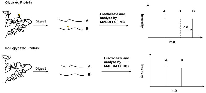
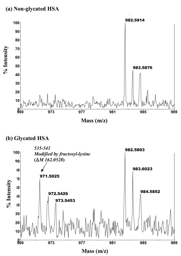
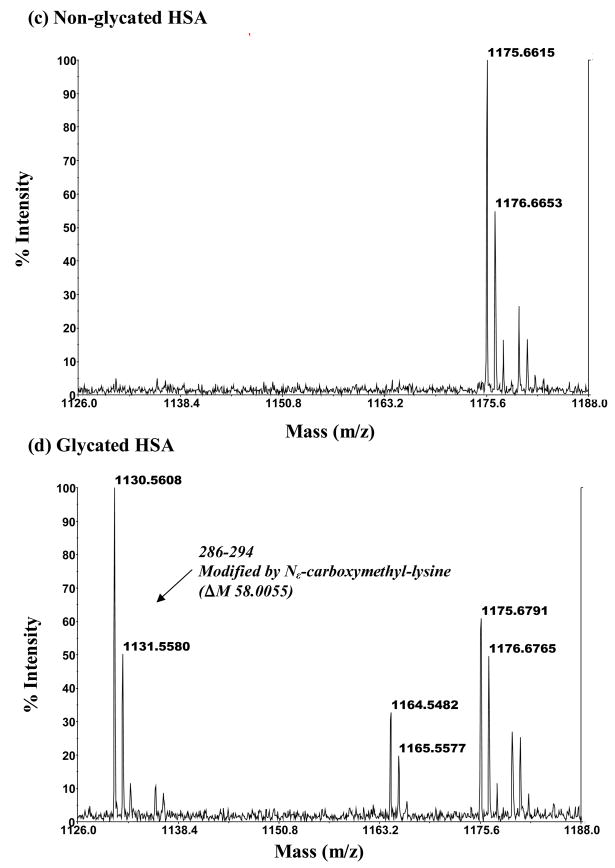
Methods: Minimally-glycated and normal HSA (used as a control) were digested with trypsin, Glu-C or Lys-C, followed by fractionation of the resulting peptides and their analysis by matrix-assisted laser desorption/ionization time-of-flight mass spectrometry (MALDI-TOF MS) to determine the structures and locations of glycation adducts.

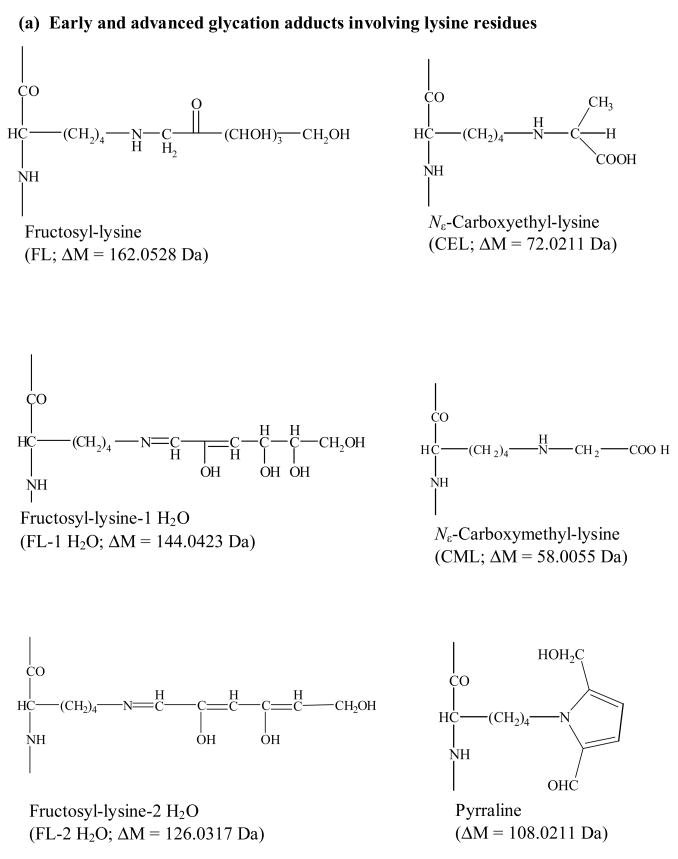
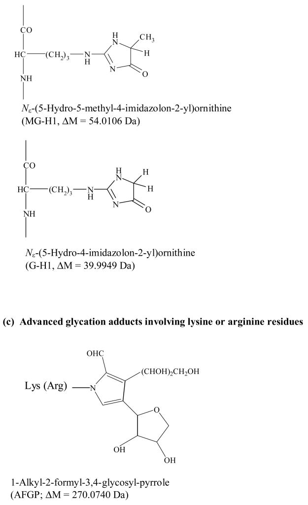
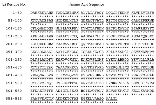
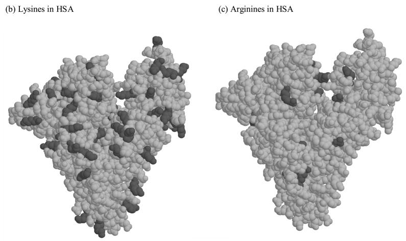
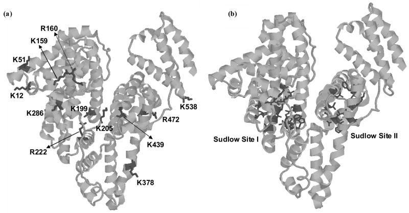
Results: Several specific lysine and arginine residues were identified as modification sites in minimally-glycated HSA. Residues K12, K51, K199, K205, K439 and K538 were found to be modified through the formation of fructosyl-lysine, while the modification of K159 and K286 involved the formation of pyrraline or N(epsilon)-carboxymethyl-lysine, respectively. Lysine K378 was found to give N(epsilon)-carboxyethyl-lysine in some forms of glycated HSA but fructosyl-lysine in other forms. Residues R160 and R472 produced a modification based on N(epsilon)-(5-hydro-4-imidazolon-2-yl)ornithine. Lysine R222 was modified to produce argpyrimidine, N(epsilon)-[5-(2,3,4-trihydroxybutyl)-5-hydro-4-imidazolon-2-yl]ornithine or tetrahydropyrimidine.

Conclusions: With the exception of K12, K199, K378, K439 and K525, all of the observed sites of modification for minimally-glycated HSA were new to this current study. The fact that many of these glycation-related modifications are located at or near known drug binding sites on HSA explains why some differences have been previously noted in the binding of certain drugs to normal vs glycated HSA.


Figures

Figure 1
Figure 1
Possible early and advanced glycation adducts that involve lysine residues (a), arginine residues (b) or either lysine or arginine residues (c) on a protein.
Figure 1
Figure 1
Possible early and advanced glycation adducts that involve lysine residues (a), arginine residues (b) or either lysine or arginine residues (c) on a protein.
Figure 1
Figure 1
Possible early and advanced glycation adducts that involve lysine residues (a), arginine residues (b) or either lysine or arginine residues (c) on a protein.
Figure 2
Figure 2
Use of MALDI-TOF MS and peptide mapping to study modification sites on a glycated protein.
Figure 3
Figure 3
Strategy used for identifying potential modification sites on glycated HSA
Figure 4
Figure 4
(a) Primary sequence and (b–c) crystal structure of HSA. The lysine residues (K) are given in italics in (a) and as darkened regions in (b). The arginine residues (R) are given in bold in (a) and as darkened regions in (c). The sequence in (a) shown the regions of the primary sequence that could be identified by MALDI-TOF MS for normal HSA ( ___ ) and minimally glycated HSA (***). The plots (b) and (c) are based on PDB file 1AO6.
Figure 4
Figure 4
(a) Primary sequence and (b–c) crystal structure of HSA. The lysine residues (K) are given in italics in (a) and as darkened regions in (b). The arginine residues (R) are given in bold in (a) and as darkened regions in (c). The sequence in (a) shown the regions of the primary sequence that could be identified by MALDI-TOF MS for normal HSA ( ___ ) and minimally glycated HSA (***). The plots (b) and (c) are based on PDB file 1AO6.
Figure 5
Figure 5
MALDI-TOF MS mass spectra for modified peptides found in digests of glycated HSA but absent in digests of normal HSA. The mass spectrum in (b) is for a modified peptide with a mass of 971.50 Da that originates from the 535–541 region of HSA and represents a fructosyl-lysine modification (ΔM, 162.05 Da). The mass spectrum in (d) is for a modified peptide with a mass of 1130.56 Da that corresponds to the 286–294 region of HSA and represents a Nε-carboxymethyl-lysine modification (ΔM, 58.01 Da). The mass spectra in (a) and (c) show the absence of these modified peptides in digests of normal HSA.
Figure 5
Figure 5
MALDI-TOF MS mass spectra for modified peptides found in digests of glycated HSA but absent in digests of normal HSA. The mass spectrum in (b) is for a modified peptide with a mass of 971.50 Da that originates from the 535–541 region of HSA and represents a fructosyl-lysine modification (ΔM, 162.05 Da). The mass spectrum in (d) is for a modified peptide with a mass of 1130.56 Da that corresponds to the 286–294 region of HSA and represents a Nε-carboxymethyl-lysine modification (ΔM, 58.01 Da). The mass spectra in (a) and (c) show the absence of these modified peptides in digests of normal HSA.
Figure 6
Figure 6
The 3-dimensional structure of HSA, showing (a) the sites of both early and advanced glycation adducts on HSA and (b) the locations of Sudlow sites I and II. These structures are based on PDB file 1AO6.

References

    1. Theodore Peters J, editor. All about albumin: biochemistry, genetics, and medical applications. San Diego, CA: Academic Press Limited; 1996.
    1. Muller WA, Fehske KJ, Schlafer SAC. Structure of binding sites on albumin. In: Reidenberg MM, Erill S, editors. Drug-protein binding. New York: Praeger Publishers; 1986.
    1. Sjoholm I. The specificity of drug binding sites on human serum albumin. In: Reidenberg MM, Erill S, editors. Drug-protein binding. New York: Praeger Publishers; 1986.
    1. He XM, Carter DC. Structure of human serum albumin. Science. 1990;249:302–303. - PubMed
    1. Gwilt PR, Nahhas RR, Tracewell WG. The effects of diabetes mellitus on pharmacokinetics and pharmacodynamics in humans. Clin Pharmacokinet. 1991;20:477–490. - PubMed

Publication types

MeSH terms

LinkOut - more resources