. 2007 Oct;36(2):211-21.
doi: 10.1016/j.mcn.2007.06.008. Epub 2007 Jul 24.

The Drosophila ARC homolog regulates behavioral responses to starvation

Affiliations

The Drosophila ARC homolog regulates behavioral responses to starvation

Mark D Mattaliano et al. Mol Cell Neurosci. 2007 Oct.

Abstract

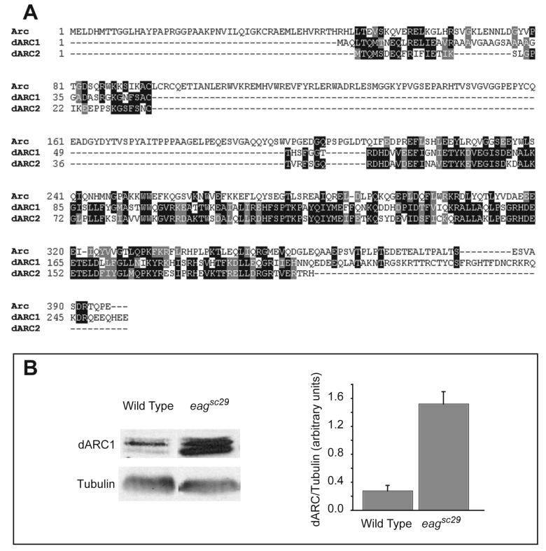
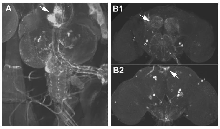
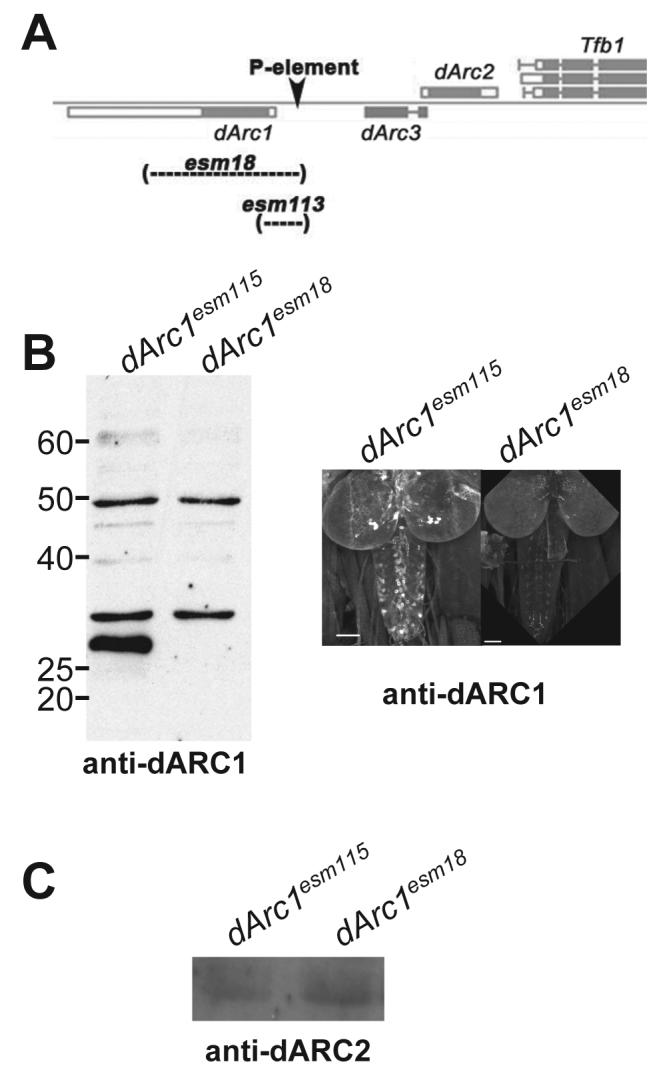
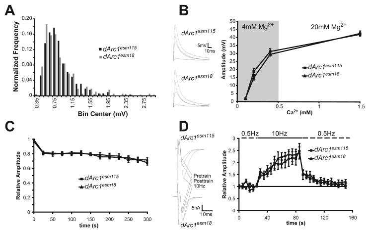
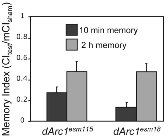
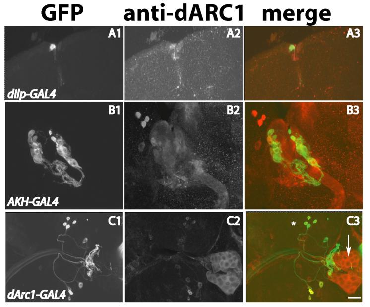
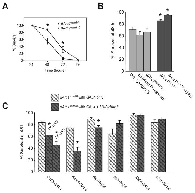
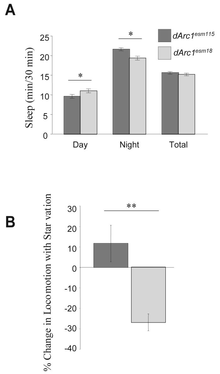
The gene encoding dARC1, one of three Drosophila homologs of mammalian activity-regulated cytoskeleton-associated protein (ARC), is upregulated in both seizure and muscular hypercontraction mutants. In this study we generate a null mutant for dArc1 and show that this gene is not involved in synaptic plasticity at the larval neuromuscular junction or in formation or decay of short-term memory of courtship conditioning, but rather is a modifier of stress-induced behavior. dARC1 is expressed in a number of neurosecretory cells and mutants are starvation-resistant, exhibiting an increased time of survival in the absence of food. Starvation resistance is likely due to the fact that dArc1 mutants lack the normal hyperlocomotor response to starvation, which is almost universal in the animal kingdom. dARC1 acts in insulin-producing neurons of the pars intercerebralis to control this behavior, but does not appear to be a general regulator of insulin signaling. This suggests that there are multiple modes of communication between the pars and the ring gland that control starvation-induced behavioral responses.


Figures

Figure 1
Figure 1. Drosophila has Arc homologs
A) Alignment of rat ARC with the Drosophila dARC1 and dARC2 proteins. The fly proteins are smaller and homologous primarily to the C-terminus of the mammalian protein. B) Drosophila dARC1 is induced by neuronal hyperexcitability. Immunoblots of dARC1 and tubulin (as a loading control) from wild type Canton S and eagsc29 heads are shown at left. Quantification of four experiments shows that dARC1 is significantly (P < 0.001, Student's T-test) induced by the seizure activity of the eag potassium channel mutant.
Figure 2
Figure 2. dARC1 expression in larval and adult CNS
Dissected larvae or adult brains were stained with rabbit anti-dARC1 (1:1000) and imaged using confocal microscopy. A) Third instar larval dissection. Several pairs of large neurons are seen in the central brain as well as in the ventral ganglion. Arrow indicates the larval ring gland. B) Adult brain. Top panel is an anterior confocal stack. Arrow indicates antennal lobes. Bottom is a posterior view showing large paired central brain neurons. Arrow indicates the pars intercerebralis.
Figure 3
Figure 3. Generation of a null mutation in dArc1
A) Schematic map of the dArc region showing exon structure. Transcription units that go left to right are above the line, while units that go right to left are below. A P-element inserted between dArc1 and dArc3 (position indicated by arrowhead) was mobilized to generation precise excisions (control lines dArc1esm115 and dArc1esm295) and deletions that take out all (dArc1esm18) or part (dArc1esm113) of the dArc1 locus. Deleted regions are indicated by a dotted line. B) dArc1esm18 is a null allele. Extracts of mutant and precise excision flies were immunoblotted with anti-dARC1 (left) and third instar larval brains of the same genotypes were stained (right). A protein of the predicted molecular weight for dARC1 is missing in the mutant flies, while high molecular weight background bands remain. Mutant brains lack staining in central brain and in the region of pars neurosecretory cells, but show some residual background staining in the ventral ganglion. Scale bar = 50 μm. C) Deletion of dArc1 does not significantly affect levels of dARC2. Head extracts from deletion and precise excision flies were immunoblotted with anti-dARC2. Both lines show normal dARC2 expression.
Figure 4
Figure 4. Mutation of dArc1 does not alter basal transmission or short-term synaptic plasticity at the larval neuromuscular junction
Recordings were made from muscle 6 from abdominal segments A3-5 in HL3 with indicated ion concentrations. A) Miniature EJPs were recorded from mutant and precise excision controls in 1.5 mM calcium with 3 μM TTX. No difference was seen in either frequency or amplitude of mEJPs. B) Evoked EJPs were recorded in mutant and precise excision animals in varying calcium. Higher calcium levels required elevation of magnesium to suppress muscle contraction. Averages of 10 traces from each calcium concentration are shown at left. Amplitude increased as a function of extracellular calcium, but did not differ between genotypes. C) High frequency stimulation does not differentially alter vesicle recycling in mutant and precise excision controls. 10 Hz stimulation in 1.5 mM calcium and 20 mM magnesium reduced release by about 30% in both genotypes measured in current clamp. D) Post-tetanic potentiation is identical in mutant and precise excision control animals as measured in voltage clamp. Averages of 10 traces from the 0.5 Hz pre-tetanus test pulses, the 10 Hz tetanus, and the post-tetanus 0.5 Hz test pulses are shown at left.
Figure 5
Figure 5. Mutation of dArc1 does not disrupt learning or short-term memory of courtship conditioning
Male flies were exposed to a mated female for one hour. Behavior during the training period was indistinguishable between the null dArc1esm18 and precise excision control dArc1esm115 flies (data not shown). Sham trained (1 h in an empty chamber) controls were done for each genotype. Memory was assessed at 10 min and 2 h after training by measuring courtship of a virgin female. Data are expressed as CItest/meanCIsham where a value of 1 indicates no memory. Both genotypes have normal initial memory and normal decay.
Figure 6
Figure 6. dARC1 expression in neuroendocrine cells
Dissected larvae or adult brains were stained with rabbit anti-dARC1 (1:1000) and imaged using confocal microscopy. Scale bar = 40 μ. A) Adult brain from a dilp-GAL4/+; UAS-mCD8GFP/+ animal. Panel A1 shows GFP expression in a subset of dilp+ pars neurons. Panel A2 shows anti-dARC1 staining. Panel A3 is an overlay showing a subset of the dARC1+ cells are also dilp+. B) Larval brain from an AKHGAL4/+; UAS-mCD8GFP/+ animal. Panel B1 shows GFP expression in AKH cells in the ring gland. Panel B2 shows anti-dARC1 staining. Panel B3 is an overlay showing the AKH-GAL4 cells are dARC1+. C) Larval brain from a dArc1-GAL4/+; UAS-mCD8GFP/+ animal. Panel C1 shows GFP expression in neurons innervating the ring gland. Panel C2 shows anti-dARC1 staining of both neurons and ring gland. Panel C3 is an overlay showing the neuronal subset of dARC1+ cells (indicated by *) which have dArc1-GAL4 expression. Arrow indicates the larval ring gland.
Figure 7
Figure 7. dArc1 mutants are resistant to starvation
1-3 day old animals were placed in vials containing 1% agar in water in a 25°C, 70% humidity environment room. A) Time course of death from starvation. dArc1esm18 animals show significantly greater survival than WT precise excision dArc1esm115 flies at 48 and 72 h after withdrawal from food. B) Null mutants, with or without the UAS-dArc1 transgene (dark bars) are significantly more resistant to starvation at 48 h compared with Canton S wildtype, the starting P element line or a precise excision control. C) The starvation resistance of dArc1esm18 can be reversed by expression of dARC1 in selected neuronal populations. Panneural expression with C155-GAL4 gives dose-dependent effects, comparing 0, 1 or 2 copies of the UAS-dArc1 transgene. Driving UAS-dArc1 with the dArc1 promoter or dilp-GAL4 significantly decreases survival. Expression in AKH- or Amn-expressing cells does not significantly change survival. In all panels, * indicates P < 0.05, ANOVA with Fisher's PLSD post-hoc analysis.
Figure 8
Figure 8. dArc1 mutants have alterations in basal and starvation-modulated locomotion
Data from dArc1esm18 are represented by dark gray bars; data from dArc1esm115 are indicated by light gray bars. A) dArc1esm18 animals have altered sleep patterns. Locomotor activity of females of the indicated genotype were collected in a Trikinetics monitor (Waltham, MA) in a 12 h light:dark cycle. Sleep was measured as bouts of 5 min of uninterrupted inactivity, as previously described (Shaw et al., 2000). Both day and night sleep differed between mutant and revertant (* indicates P < 0.05, ANOVA with Tukey-Kramer post hoc test; n > 45). Total sleep was not significantly different between genotypes. B) dArc1esm18 animals have an abnormal locomotor response to starvation. Locomotor activity was averaged for the time window 12-15 h after the initiation of starvation and compared to the average activity of fed flies. The % change from fed flies is shown. dArc1esm18 animals show a significant decrease in locomotor activity with starvation compared to revertant controls (** indicates P < 0.001, Student's t-test, n ≥ 115 for each genotype).

References

    1. Bader MF, Doussau F, Chasserot-Golaz S, Vitale N, Gasman S. Coupling actin and membrane dynamics during calcium-regulated exocytosis: a role for Rho and ARF GTPases. Biochim Biophys Acta. 2004;1742:37–49. - PubMed
    1. Berasi SP, Huard C, Li D, Shih HH, Sun Y, Zhong W, Paulsen JE, Brown EL, Gimeno RE, Martinez RV. Inhibition of gluconeogenesis through transcriptional activation of EGR1 and DUSP4 by AMP-activated kinase. J Biol Chem. 2006;281:27167–27177. - PubMed
    1. Beumont PJ, Arthur B, Russell JD, Touyz SW. Excessive physical activity in dieting disorder patients: proposals for a supervised exercise program. The International journal of eating disorders. 1994;15:21–36. - PubMed
    1. Bohni R, Riesgo-Escovar J, Oldham S, Brogiolo W, Stocker H, Andruss BF, Beckingham K, Hafen E. Autonomous control of cell and organ size by CHICO, a Drosophila homolog of vertebrate IRS1-4. Cell. 1999;97:865–875. - PubMed
    1. Broughton SJ, Piper MD, Ikeya T, Bass TM, Jacobson J, Driege Y, Martinez P, Hafen E, Withers DJ, Leevers SJ, Partridge L. Longer lifespan, altered metabolism, and stress resistance in Drosophila from ablation of cells making insulin-like ligands. Proc Natl Acad Sci U S A. 2005;102:3105–3110. - PMC - PubMed

Publication types

MeSH terms