. 2007;35(17):5646-57.
doi: 10.1093/nar/gkm629. Epub 2007 Aug 20.

Real-time assembly and disassembly of human RAD51 filaments on individual DNA molecules

Affiliations

Real-time assembly and disassembly of human RAD51 filaments on individual DNA molecules

Thijn van der Heijden et al. Nucleic Acids Res. 2007.

Abstract

The human DNA repair protein RAD51 is the crucial component of helical nucleoprotein filaments that drive homologous recombination. The molecular mechanistic details of how this structure facilitates the requisite DNA strand rearrangements are not known but must involve dynamic interactions between RAD51 and DNA. Here, we report the real-time kinetics of human RAD51 filament assembly and disassembly on individual molecules of both single- and double-stranded DNA, as measured using magnetic tweezers. The relative rates of nucleation and filament extension are such that the observed filament formation consists of multiple nucleation events that are in competition with each other. For varying concentration of RAD51, a Hill coefficient of 4.3 +/- 0.5 is obtained for both nucleation and filament extension, indicating binding to dsDNA with a binding unit consisting of multiple (> or =4) RAD51 monomers. We report Monte Carlo simulations that fit the (dis)assembly data very well. The results show that, surprisingly, human RAD51 does not form long continuous filaments on DNA. Instead each nucleoprotein filament consists of a string of many small filament patches that are only a few tens of monomers long. The high flexibility and dynamic nature of this arrangement is likely to facilitate strand exchange.


Figures

Figure 1.
Figure 1.
(A) Schematic drawing of the magnetic tweezers setup. A DNA molecule is attached at one end to the bottom of the flow cell and at the other end to a magnetic bead. This molecule can be stretched using a pair of magnets placed above the flow cell. The bead position and thus the end-to-end distance of the DNA molecule is determined using video microscopy and image analysis. In such a setup, the interaction of RAD51 with DNA can be followed in real time because binding induces a change in end-to-end distance of the tethered molecule. (B) The tethered molecule in the setup is characterized using force-extension measurements to confirm a correct contour and persistence length. The force-extension behavior of dsDNA is fit using the worm-like chain model (red line) yielding a contour length of 2.9 ± 0.1 µm and a persistence length of 49 ± 3 nm, whereas ssDNA displays a different flexibility behavior (black line as a guide to the eye) which cannot be described by worm-like chain (45). After filament formation in the presence of Ca2+, the DNA molecule is elongated and stiffened yielding a contour length of 4.0 ± 0.1 µm and a persistence length of 268 ± 17 nm (blue line) for this particular case.
Figure 2.
Figure 2.
RAD51 filament assembly and disassembly on dsDNA. (A) An 8 kb dsDNA molecule extends by 45% after flushing in 187 nM RAD51 in the presence of 25 mM Tris–HCl (pH 7.5), 5 mM MgCl2, 25 mM KCl, 1 mM DTT and 1 mM ATP. (B) After removal of free RAD51 in solution, disassembly of a RAD51-DNA filament is observed in the presence of Mg2+. (C) In the presence of Ca2+ instead of Mg2+, a binding behavior similar to that of Mg2+ is observed. (D) In the presence of Ca2+, however, disassembly is not observed and the end-to-end distance remained unaffected upon the removal of free RAD51 in solution. The solid lines in the figures show the result of Monte Carlo simulations using a rate for nucleation and filament extension to describe filament assembly. For the disassembly reaction, a single disassembly rate is taken into account. Solid lines in panels A and B are fits, whereas the line in panel C is plotted without any adjustable parameters. Dashed horizontal lines indicate the DNA end-to-end length in the absence of RAD51. Reaction rates were determined from the average of multiple experiments for each condition (n ∼ 10).
Figure 3.
Figure 3.
RAD51 filament assembly and disassembly on ssDNA (A) In the presence of Mg2+ and 1 mM ATP, 187 nM RAD51 starts extending an 8.6 knt ssDNA molecule directly after the flush is stopped. In contrast to dsDNA (Figure 2C), the observed extension was slow and very irregular, as emphasized in the inset. The blue line is a fit based on the Monte Carlo simulations allowing disassembly per filament end. Nucleation and growth rates where taken from the Ca2+ data (see subsequently). (B) After removal of free RAD51 in solution, the DNA end-to-end distance decreased, indicating disassembly of the RAD51-ssDNA filament. The solid line is a fit with a double exponential (see text). (C) In the presence of Ca2+, the observed growth profile is similar to the one observed on dsDNA. The solid lines are fits based on the Monte Carlo simulations allowing only nucleation and growth. (D) After removal of free RAD51 in solution, the end-to-end distance remained constant when Ca2+ is present. Dashed horizontal lines indicate the DNA end-to-end length in the absence of RAD51. Reaction rates were determined from the average of multiple experiments for each condition (n ∼ 10).
Figure 4.
Figure 4.
Results from Monte Carlo simulations for the binding of a recombinase on DNA for different levels of cooperativity. If only random binding (nucleation) occurs along the contour length of the DNA molecule (non-cooperative binding, see top left), an exponential growth profile is obtained (black line). The obtained elongation is smaller than 50% due to the presence of nucleotide positions on the DNA where no further binding can occur. However, if filament extension is fast compared to nucleation, e.g. for a ratio larger than 106 (strong cooperative binding, see bottom right), the growth profile becomes linear and the molecule can be fully covered by the protein up to the maximum 50% elongation (blue line). For intermediate ratios between filament extension and nucleation, sigmoid-like shaped growth profiles are observed (green, red, gray lines).
Figure 5.
Figure 5.
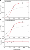
Concentration-dependent RAD51 filament assembly rates on dsDNA in the presence of Ca2+ and 1 mM ATP. (A) Nucleation rate. The nucleation data was fit with the Hill equation, yielding a Hill coefficient n = 4.34 ± 0.56, a substrate concentration where half-maximal activity occurs S0.5 = 149 ± 4 nM, and a maximum nucleation rate ki,max = (0.61 ± 0.03)×10−3 s−1 bp−1. (B) Extension rate per filament patch. The extension rate per filament patch was fit with a Hill coefficient n = 4.35 ± 0.46, S0.5 = 149 ± 4 nM, and ki,max = 0.150 ± 0.006 pentamers s−1 end−1. (C) The ratio between extension per filament patch and nucleation is found to remain constant as a function of [RAD51]. The dotted lines correspond to binding rates that follow a Michaelis–Menten relation, indicative of monomeric nucleation and extension per filament patch. This clearly fails to fit the data.
Figure 6.
Figure 6.
(A) Snapshots of RAD51-DNA filament patches at different times during a Monte Carlo simulation in the absence of dissociation. This simulation is carried out for knucl = 5 × 10⁻⁷ site−1 (Monte Carlo step)−1, kext = 1 × 10⁻⁴ (Monte Carlo step)−1. Due to the fast nucleation rate, multiple filaments are formed along the DNA substrate. As a RAD51 pentamer covers 15 nt or base pair upon binding, nucleation or filament extension can only occur if sufficient space is available. In the bottom panel, the simulation has reached its final state since no further pentamers can bind. (B) Filament-length distribution in the final state for dsDNA in the presence of Mg2+, ssDNA with Mg2+ and DNA with Ca2+. The distributions for ss- and dsDNA with Ca2+ are practically identical. (C) Final elongation of a RAD51-DNA filament as a function of the ratio between rates for filament extension and nucleation. For low kext/knucl, a plateau of 38% extension is obtained, whereas for high kext/knucl the full 50% extension can be reached. The open circles represent the elongations of ss- and dsDNA by RAD51 obtained in the presence of Ca2+ in our measurements, and the elongation by RecA on dsDNA taken from Ref. (11).
Figure 7.
Figure 7.
(A) Growth curves from Monte Carlo simulations (solid lines) at different ratios between filament extension and nucleation fitted to an experimental binding profile of RAD51 on ssDNA in the presence of Ca2+ (gray circles). Inset shows the least-squares-fit residuals obtained for several ratios of kext/knucl. As can be seen in the inset, the best fit is obtained at a ratio of 220:1, where the least-squares-fit residuals are minimal. The minimum is determined using a parabola. (B) Monte Carlo simulations of RAD51 filament formation on ssDNA allowing disassembly during growth per monomer (black) or per filament end (blue), without changing the rates for nucleation and growth.

Similar articles

Cited by

References

    1. West SC. Molecular views of recombination proteins and their control. Nat. Rev. Mol. Cell Biol. 2003;4:435–445. - PubMed
    1. Wyman C, Kanaar R. Homologous recombination: Down to the wire. Curr. Biol. 2004;14:R629–R631. - PubMed
    1. Conway AB, Lynch TW, Zhang Y, Fortin GS, Fung CW, Symington LS, Rice PA. Crystal structure of a Rad51 filament. Nat. Struct. Mol. Biol. 2004;11:791–796. - PubMed
    1. Yu X, Jacobs SA, West SC, Ogawa T, Egelman EH. Domain structure and dynamics in the helical filaments formed by RecA and Rad51 on DNA. Proc. Natl Acad. Sci. USA. 2001;98:8419–8424. .S. - PMC - PubMed
    1. Yu X, VanLoock MS, Yang S, Reese JT, Egelman EH. What is the structure of the RecA-DNA filament? Curr. Protein Pept. Sc. 2004;5:73–79. - PubMed

Publication types