. 2007 Oct;27(20):7248-65.
doi: 10.1128/MCB.02199-06. Epub 2007 Aug 20.

pRb-dependent cyclin D3 protein stabilization is required for myogenic differentiation

Affiliations

pRb-dependent cyclin D3 protein stabilization is required for myogenic differentiation

Francesca De Santa et al. Mol Cell Biol. 2007 Oct.

Abstract

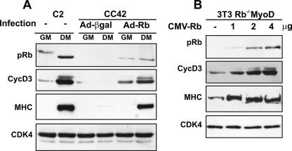
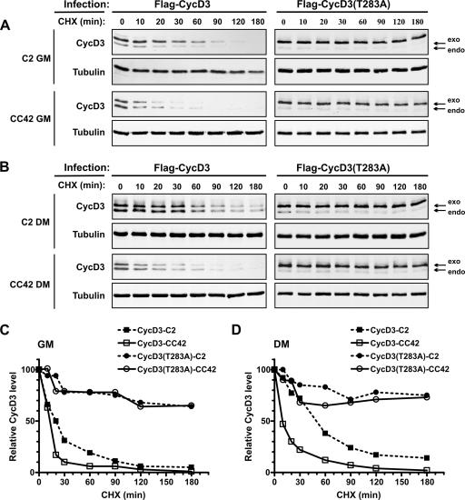
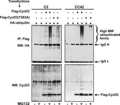
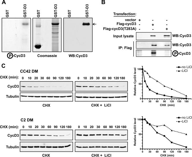
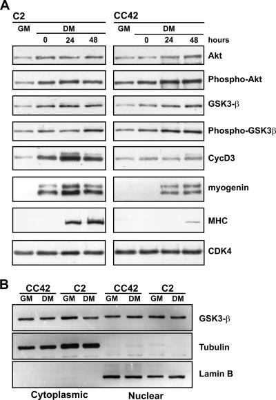
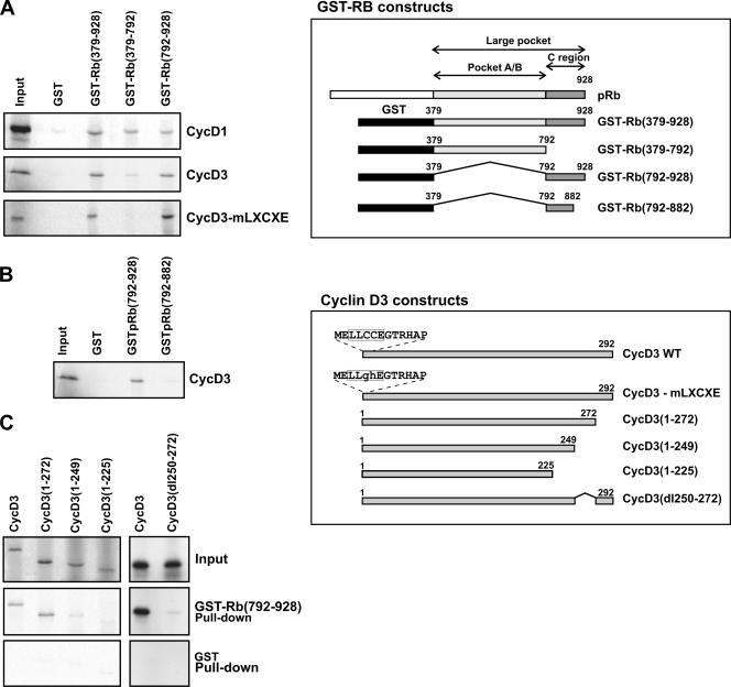
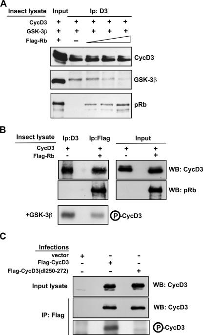
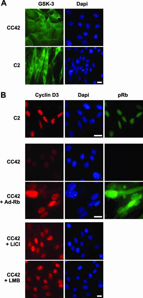
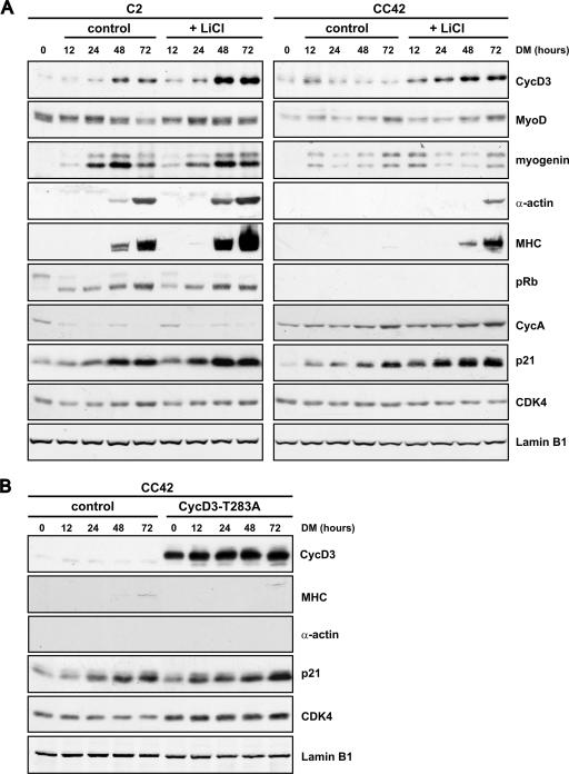
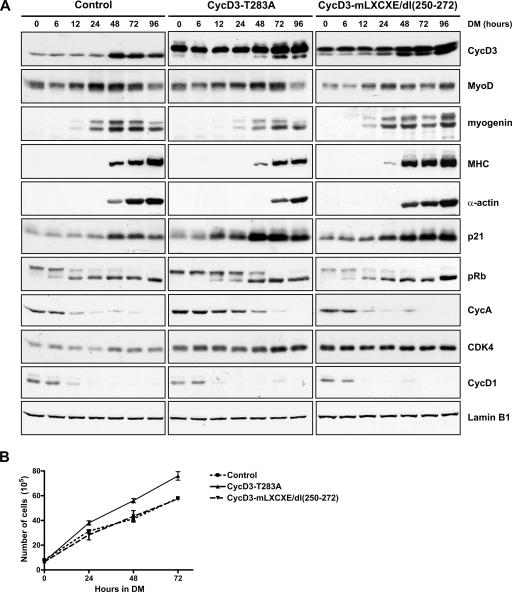
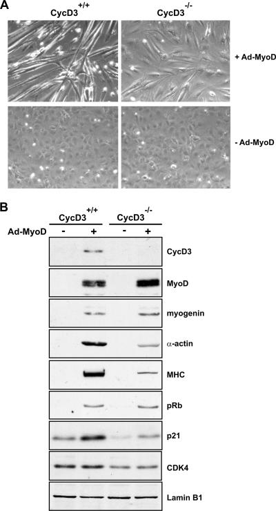
The expression of retinoblastoma (pRb) and cyclin D3 proteins is highly induced during the process of skeletal myoblast differentiation. We have previously shown that cyclin D3 is nearly totally associated with hypophosphorylated pRb in differentiated myotubes, whereas Rb-/- myocytes fail to accumulate the cyclin D3 protein despite normal induction of cyclin D3 mRNA. Here we report that pRb promotes cyclin D3 protein accumulation in differentiating myoblasts by preventing cyclin D3 degradation. We show that cyclin D3 displays rapid turnover in proliferating myoblasts, which is positively regulated through glycogen synthase kinase 3beta (GSK-3beta)-mediated phosphorylation of cyclin D3 on Thr-283. We describe a novel interaction between pRb and cyclin D3 that maps to the C terminus of pRb and to a region of cyclin D3 proximal to the Thr-283 residue and provide evidence that the pRb-cyclin D3 complex formation in terminally differentiated myotubes hinders the access of GSK-3beta to cyclin D3, thus inhibiting Thr-283 phosphorylation. Interestingly, we observed that the ectopic expression of a stabilized cyclin D3 mutant in C2 myoblasts enhances muscle-specific gene expression; conversely, cyclin D3-null embryonic fibroblasts display impaired MyoD-induced myogenic differentiation. These results indicate that the pRb-dependent accumulation of cyclin D3 is functionally relevant to the process of skeletal muscle cell differentiation.


Figures

FIG. 1.
FIG. 1.
Ectopic expression of pRb restores normal levels of cyclin D3 protein in differentiating Rb/ myocytes. (A) Proliferating CC42 myoblasts (Rb/) were infected with Adeno-Rb or Adeno-β-gal and cultured in GM or DM for 48 h. Equal amounts of protein from cell lysates were analyzed by immunoblotting with antibodies specific to pRb, cyclin D3, or MHC. As positive control, C2 myoblasts (Rb+/+) cultured in GM or in DM were analyzed in parallel. Equal loading of the extracts was monitored by immunoblotting with anti-cdk4. (B) Proliferating 3T3 Rb/ fibroblasts were infected with the pBABEpuro-MyoD retrovirus and selected for 3 days with puromycin. The puromycin-resistant cell population (3T3 Rb−/−MyoD) was then transfected with increasing amounts of the CMV-Rb expression construct; the CMV-Neo-Bam empty vector was included in transfection mixtures to normalize for DNA amount. Cells were transferred to DM 24 h posttransfection and harvested after 48 h. Equal amounts of protein from cell lysates were analyzed for pRb, cyclin D3, or MHC protein expression by immunoblotting.
FIG. 2.
FIG. 2.
Kinetics of cyclin D3 or cyclin D3(T283A) protein turnover. C2 (Rb+/+) or CC42 (Rb/) myoblasts were infected with retroviruses encoding wild-type Flag-CycD3 or Flag-CycD3(T283A). The infected cells were cultured in GM or in DM for 48 h and then treated with CHX (50 μg/ml) over a 180-min time course. Cells were harvested at the indicated time points after the addition of CHX, and whole-cell extracts were prepared. Amounts of 60 μg of protein from cells infected with Flag-CycD3 or 30 μg from those infected with Flag-CycD3(T283A) were analyzed by Western blotting. (A) Immunoblot of cyclin D3 and α-tubulin expression by proliferating C2 or CC42 myoblasts. (B) Immunoblot of cyclin D3 and α-tubulin expression by differentiated C2 or CC42 myocytes. The arrows indicate the endogenous and the exogenous Flag-tagged cyclin D3. The blots shown are representative of the results of three independent experiments. (C, D) The immunoblots in panels A and B were detected by using an Odyssey infrared imaging system (Li-Cor), and the intensities of the protein bands were quantitated. The values for cyclin D3 were normalized to those for α-tubulin and are presented as the percentage of the amount of normalized cyclin D3 in cells not treated with CHX (time, 0 min).
FIG. 3.
FIG. 3.
Thr-283 is required for cyclin D3 ubiquitination. C2 (Rb+/+) or CC42 (Rb/) proliferating myoblasts were transfected with expression vectors encoding wild-type Flag-CycD3 or Flag-CycD3(T283A) together with an expression vector for HA-tagged ubiquitin. Transfected cells were treated with the proteasome inhibitor MG132 (10 μM) or with the solvent dimethyl sulfoxide for 4 h before harvesting. Cell extracts were immunoprecipitated with anti-Flag antibody. Immunocomplexes were resolved by SDS-PAGE and analyzed by using either anti-HA (upper panels) or anti-cyclin D3 (lower panels) antibodies. MW, molecular weight; IgG H, immunoglobulin G heavy chains; IgG L, immunoglobulin G light chains; WB, Western blot; IP, immunoprecipitate; +, present; −, absent.
FIG. 4.
FIG. 4.
GSK-3β phosphorylates cyclin D3 on Thr-283. (A) Glutathione-Sepharose-bound GST or GST-D3 (1 μg) was incubated with human recombinant GSK-3β in a kinase assay mixture containing [γ-32P]ATP. Proteins were resolved by SDS-PAGE, stained with Coomassie blue, dried, and subjected to autoradiography. Amounts of 250 ng of GST or GST-D3 were resolved on a parallel gel, transferred to a membrane, and immunoblotted with an antibody to cyclin D3. WB, Western blot; P-CycD3, phospho-cyclin D3. (B) NIH 3T3 fibroblasts were transfected with wild-type Flag-CycD3, Flag-CycD3(T283A), or pFlex-1 empty vector. Whole-cell extracts (500 μg) were immunoprecipitated (IP) with anti-FLAG antibody. Cyclin D3 levels were analyzed in input lysates (5%) and in the anti-Flag immunoprecipitates (20%) by immunoblotting; the remainder of the anti-Flag immunoprecipitates (80%) was incubated with human recombinant GSK-3β and [γ-32P]ATP. Proteins were resolved by SDS-PAGE, and phospho-cyclin D3 (P-CycD3) visualized by autoradiography. WB, Western blot; +, present; −, absent. (C) CC42 (Rb/) or C2 (Rb+/+) confluent myoblasts were exposed to DM for 48 h and then incubated with or without LiCl (20 mM) for 6 h and treated with CHX (50 μg/ml) for the times indicated. Equal amounts of protein from cell lysates were examined by Western blotting with antibodies against cyclin D3 and α-tubulin. The blots shown are representative of three independent experiments. The graphs on the right show the quantitation of the Western blot data by using an Odyssey infrared imaging system (Li-Cor). The values for cyclin D3 were normalized to those for α-tubulin and are presented as the percentage of the amount of normalized cyclin D3 in cells not treated with CHX (time, 0 min).
FIG. 5.
FIG. 5.
Analysis of expression, phosphorylation state, and subcellular localization of GSK-3β in Rb+/+ and Rb/ differentiating myoblasts. (A) C2 (Rb+/+) or CC42 (Rb/) myoblasts were grown to 90% confluence and then shifted to DM. Whole-cell extracts were prepared from proliferating myoblasts (GM) or from cultures exposed to DM for the indicated periods of time. Equal amounts of protein were separated by SDS-PAGE and subjected to immunoblot analysis using antibodies against Akt, GSK-3β, cyclin D3, myogenin, MHC, and cdk4 (as loading control). The phosphorylation of Akt or GSK-3β was analyzed with phospho-specific antibodies that recognize the phosphorylated Ser-473 residue of Akt or Ser-9 residue of GSK-3β. (B) C2 and CC42 myoblasts were cultured in GM or exposed to DM for 48 h. Equal amounts of protein (30 μg) from nuclear and cytoplasmic fractions were separated on denaturing gels and subjected to Western blot analysis with antibodies against GSK-3β, α-tubulin, or lamin B1.
FIG. 6.
FIG. 6.
Binding of cyclin D3 to pRb in vitro. The schematic in the upper inset shows the structure of the GST-Rb fusion proteins used and illustrates the A/B pocket region and the carboxy-terminal region of Rb. The lower inset shows schematic representations of wild-type (WT) cyclin D3 and cyclin D3 mutants carrying substitutions in the N-terminal LXCXE motif or deletions in the carboxy-terminal region. (A and B) The indicated GST-Rb fusion proteins or GST alone were bound to glutathione-Sepharose beads and incubated with in vitro-translated, 35S-labeled cyclin D1, cyclin D3, or the cyclin D3-mLXCXE mutant. Bound proteins were resolved by SDS-PAGE and processed for autoradiography. (C) The GST-Rb(792-928) fusion protein was incubated with the indicated cyclin D3 proteins translated and radiolabeled in vitro. Bound proteins were resolved by SDS-PAGE and processed for autoradiography.
FIG. 7.
FIG. 7.
pRb inhibits the association between cyclin D3 and GSK-3β, thus preventing cyclin D3 phosphorylation by GSK-3β. (A) Insect Sf9 cells were infected with baculoviruses encoding cyclin D3, GSK-3β, or Flag-Rb. Sf9 cell extract containing cyclin D3 (30 μg protein) was incubated with extract containing GSK-3β (50 μg) in the presence or absence of increasing amounts of extract containing Flag-Rb (50 μg, 150 μg, or 300 μg). Lysate from uninfected Sf9 cells was added to the reaction mixtures to adjust for total protein amount. Cyclin D3-containing complexes were then immunoprecipitated (Ip:D3). Of each immunoprecipitate, 10% was analyzed by immunoblotting with anti-cyclin D3 and 90% by immunoblotting with anti-GSK-3β or anti-Rb. The input lane contains 3 μg of protein of cyclin D3 lysate, 5 μg of GSK-3β lysate, and 5 μg of Flag-Rb lysate. The blot shown is representative of three independent experiments. (B) Insect Sf9 cells were infected with a baculovirus encoding cyclin D3 or coinfected with baculoviruses encoding cyclin D3 or Flag-Rb. The Sf9 lysate containing cyclin D3 (100 μg) was immunoprecipitated with an antibody to cyclin D3, whereas the Sf9 lysate containing cyclin D3 and Flag-Rb (100 μg) was subjected to precipitation with anti-Flag antibody. The expression of cyclin D3 and pRb was analyzed in the input lysates (5%) and in the immunoprecipitates (25%) by Western blotting. The anti-cyclin D3 (25%) and the anti-Flag (50%) immunoprecipitates were incubated in kinase reaction mixtures with human recombinant GSK-3β and [γ-32P]ATP. Samples were then subjected to SDS-PAGE and autoradiography. The results of one experiment representative of three are shown. (C) NIH 3T3 fibroblasts were infected with retroviruses expressing Flag-CycD3, Flag-CycD3-dl(250-272), or empty vector and harvested 48 h postinfection. Cell extracts (500 μg) were immunoprecipitated with anti-Flag antibody, and afterwards, 20% of each immunoprecipitate was analyzed by immunoblotting with an antibody to cyclin D3 while the remainder was incubated with human recombinant GSK-3β and [γ-32P]ATP in kinase reaction mixtures. Samples were then subjected to SDS-PAGE and autoradiography. Input lysates (5%) were also analyzed. WB, Western blot; IP, immunoprecipitate; +, present; −, absent; P-CycD3, phospho-cyclin D3.
FIG. 8.
FIG. 8.
The nuclear accumulation of cyclin D3 is restored in Rb/ myocytes by expression of exogenous pRb, inhibition of GSK-3β activity, or inhibition of nuclear export. (A) C2 or CC42 myoblasts plated on glass coverslips were exposed to DM for 48 h and then stained with a GSK-3β-specific antibody and DAPI dye. (B) C2 myoblasts exposed to DM for 48 h were stained with antibodies to cyclin D3 or pRb and DAPI dye. CC42 myoblasts plated on glass coverslips were either mock-infected or infected with Adeno-Rb and then exposed to DM for 48 h. Cells were fixed and processed for immunofluorescence with antibodies specific to cyclin D3 or pRb and DAPI dye. Where indicated, CC42 myocytes were treated with LiCl (20 mM) or leptomycin B (LMB, 40 ng/ml) for 3 h or 6 h, respectively, prior to fixation. Bar, 10 μm.
FIG. 9.
FIG. 9.
Lithium chloride (LiCl) enhances terminal differentiation of both Rb+/+ and Rb/ myocytes, but ectopic expression of a stabilized cyclin D3 mutant in Rb/ myocytes is not sufficient to reproduce the promyogenic effect of LiCl. (A) C2 or CC42 myoblasts, seeded in amounts of 2 × 10⁵ cells into 90-mm dishes, were cultured in proliferation medium for 48 h and then transferred to DM either in the presence or in the absence of LiCl at 10 mM. Cells were harvested at the indicated time points, and equal amounts of protein from whole-cell extracts were analyzed by Western blotting with antibodies to the indicated cell cycle and differentiation markers. (B) CC42 myoblasts were infected with a retrovirus expressing the cyclin D3(T283A) mutant or with the pBABEpuro control virus and selected with puromycin. Cells were seeded as described for panel A and exposed to DM after 48 h. Whole-cell extracts were prepared at the indicated times, and equal amounts of protein were analyzed by Western blotting for expression of the indicated proteins. In the experiments whose results are shown in panels A and B, equal loading was monitored by immunoblotting with anti-lamin B1. Shown are representative results from three independent experiments.
FIG. 10.
FIG. 10.
The effect of ectopic expression of stabilized cyclin D3 mutants on differentiation of Rb+/+ myoblasts. (A) C2 myoblasts were infected with pBABEpuro control virus or viruses expressing the cyclin D3(T283A) or the cyclin D3-mLXCXE/dl(250-272) mutant, both Flag-tagged at their N termini. Following selection with puromycin, cells were seeded at 2 × 10⁵ cells into 90-mm dishes in GM and transferred to DM after 48 h, at which time they reached ∼50 to 60% confluence. Whole-cell extracts were prepared at 0, 6, 12, 24, 48, 72, and 96 h after exposure to DM, and equal amounts of protein were subjected to Western blot analysis using antibodies to the indicated proteins. The blots shown are representative results of three independent experiments. (B) Growth curves in DM of C2 cells infected with cyclin D3(T283A), cyclin D3-mLXCXE/dl(250-272), or control retroviruses. Cells were seeded as described for panel A and transferred to DM after 24 h when they reached ∼15% confluence. Counts were made at the time cells were shifted to DM (time, 0) and following 24, 48, and 72 h of incubation in DM. In these culture conditions, cells reach 80 to 100% confluence on day 3, when they start fusing to form multinucleated myotubes. The curves represent the averages of the results of three experiments. Error bars show standard errors of the means.
FIG. 11.
FIG. 11.
Cyclin D3 deficiency results in defects in MyoD-induced myogenic conversion in MEFs. Wild-type and cyclin D3−/− MEFs were infected with Adeno-MyoD (+Ad-MyoD) at an MOI of 500 or mock infected (−Ad-MyoD). Cells were shifted to DM 24 h postinfection and harvested 48 h later. Phase-contrast microphotographs (A) and Western blot analyses (B) of the indicated proteins in whole-cell lysates normalized for protein amount are shown. Equal loading was verified by immunoblotting with anti-lamin B1. Shown are representative results from four independent experiments. +, present; −, absent.

Similar articles

Cited by

References

    1. Alessi, D. R., M. Andjelkovic, B. Caudwell, P. Cron, N. Morrice, P. Cohen, and B. A. Hemmings. 1996. Mechanism of activation of protein kinase B by insulin and IGF-1. EMBO J. 15:6541-6551. - PMC - PubMed
    1. Alt, J. R., J. L. Cleveland, M. Hannink, and J. A. Diehl. 2000. Phosphorylation-dependent regulation of cyclin D1 nuclear export and cyclin D1-dependent cellular transformation. Genes Dev. 14:3102-3114. - PMC - PubMed
    1. Bartkova, J., J. Lukas, M. Strauss, and J. Bartek. 1998. Cyclin D3: requirement for G1/S transition and high abundance in quiescent tissues suggest a dual role in proliferation and differentiation. Oncogene 17:1027-1037. - PubMed
    1. Berkes, C. A., and S. J. Tapscott. 2005. MyoD and the transcriptional control of myogenesis. Semin. Cell Dev. Biol. 16:585-595. - PubMed
    1. Camarda, G., F. Siepi, D. Pajalunga, C. Bernardini, R. Rossi, A. Montecucco, E. Meccia, and M. Crescenzi. 2004. A pRb-independent mechanism preserves the postmitotic state in terminally differentiated skeletal muscle cells. J. Cell Biol. 167:417-423. - PMC - PubMed

Publication types

Grants and funding