. 2007 Nov;75(11):5095-104.
doi: 10.1128/IAI.00075-07. Epub 2007 Aug 20.

Two domains of cytotoxic necrotizing factor type 1 bind the cellular receptor, laminin receptor precursor protein

Affiliations

Two domains of cytotoxic necrotizing factor type 1 bind the cellular receptor, laminin receptor precursor protein

Beth A McNichol et al. Infect Immun. 2007 Nov.

Abstract

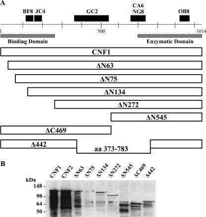
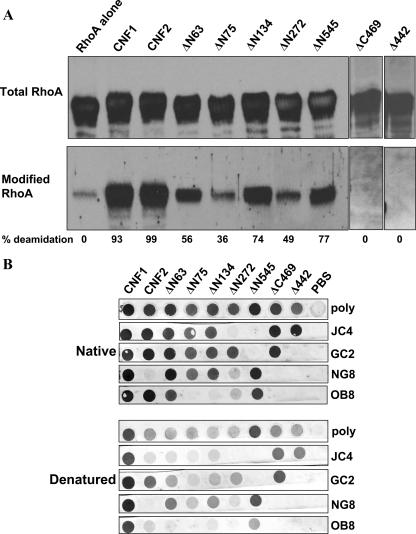
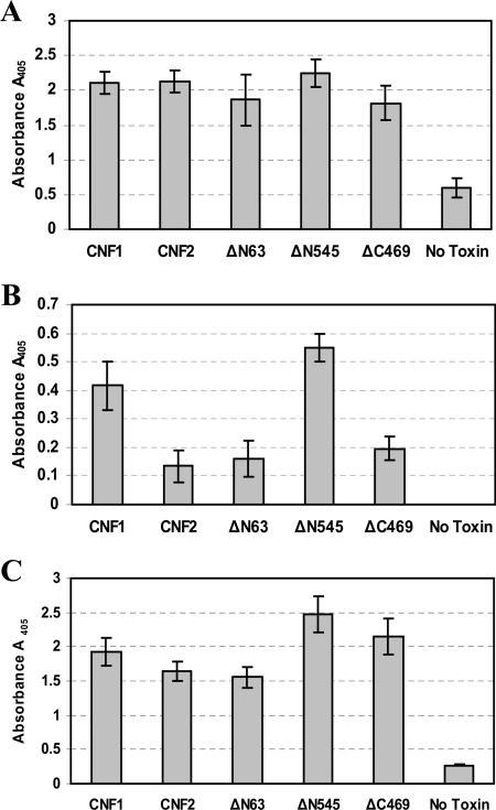
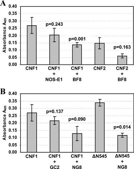
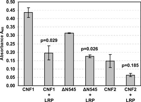
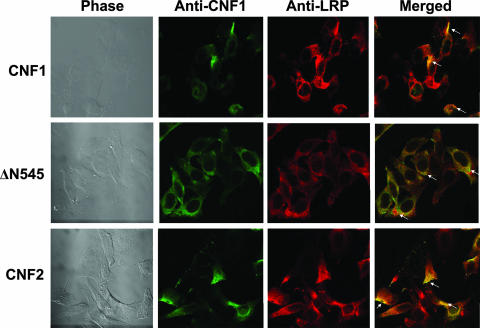
Cytotoxic necrotizing factor type 1 (CNF1) and CNF2 are highly homologous toxins that are produced by certain pathogenic strains of Escherichia coli. These 1,014-amino-acid toxins catalyze the deamidation of a specific glutamine residue in RhoA, Rac1, and Cdc42 and consist of a putative N-terminal binding domain, a transmembrane region, and a C-terminal catalytic domain. To define the regions of CNF1 that are responsible for binding of the toxin to its cellular receptor, the laminin receptor precursor protein (LRP), a series of CNF1 truncated toxins were characterized and assessed for toxin binding. In particular, three truncated toxins, DeltaN63, DeltaN545, and DeltaC469, retained conformational integrity and in vitro enzymatic activity and were immunologically reactive against a panel of anti-CNF1 monoclonal antibodies (MAbs). Based on a comparison of these truncated toxins with wild-type CNF1 and CNF2 in LRP and HEp-2 cell binding assays and in MAb and LRP competitive binding inhibition assays and based on the results of confocal microscopy, we concluded that CNF1 contains two major binding regions: one located within the N terminus, which contained amino acids 135 to 164, and one which resided in the C terminus and included amino acids 683 to 730. The data further indicate that CNF1 can bind to an additional receptor(s) on HEp-2 cells and that LRP can also serve as a cellular receptor for CNF2.


Figures

FIG. 1.
FIG. 1.
(A) Schematic diagram of CNF1 truncated toxins, with the regions and epitopes recognized by several CNF MAbs indicated at the top. MAbs JC4 and NG8 can neutralize only CNF1, while MAb BF8 can neutralize both CNF1 and CNF2. aa, amino acids. (B) Western blots of purified wild-type and truncated CNF1 toxins. Toxins (2 μg of CNF2 and the CNF1 truncated toxins and 500 ng of CNF1) were applied to SDS-PAGE gels and transferred to nitrocellulose membranes. Blots were probed with anti-CNF1 polyclonal antisera, followed by an HRP-conjugated secondary antibody and the ECL substrate. Circles indicate the position of each full-length toxin.
FIG. 2.
FIG. 2.
Enzymatic activity and conformation of CNF1 truncated toxins. (A) Western blot analysis of RhoA incubated in the presence or absence of purified toxins. Membranes were probed with a MAb against RhoA (top panel) or with antisera that recognize only the deamidated form of the GTPase (bottom panel), and reactive proteins were detected with appropriate HRP-conjugated secondary antibodies and the ECL substrate. The pixel density of total and modified RhoA was used to calculate the percent modification, and the results are indicated below the blots. Background values for the RhoA control were subtracted from each of the sample values. (B) Purified toxins (5 μg/well) were either applied directly to nitrocellulose membranes (upper panels) or denatured (with SDS, dithiothreitol, and boiling) prior to application (lower panels). As indicated on the right, blots were probed with either CNF1 polyclonal antisera (poly) or a panel of CNF1 MAbs, and reactive proteins were visualized with HRP-conjugated secondary antibodies and the diaminobenzamidine substrate.
FIG. 3.
FIG. 3.
HEp-2 and LRP receptor binding ELISAs. (A) Indirect ELISA that served as an internal control for subsequent binding ELISAs. Purified toxins (5 μg/well) were used as coating antigens and detected with CNF1 polyclonal antisera at a dilution (1:5,000) that allowed comparable signals from all toxins assayed. The data are the averages of two experiments done in triplicate. (B) HEp-2 receptor binding ELISA in which purified toxins (5 μg/well) were added to fixed HEp-2 cells for 1 h and bound toxin was detected with anti-CNF1 polyclonal sera. The data are the averages of two experiments done in triplicate. (C) LRP binding ELISA in which purified toxins were incubated in LRP-coated ELISA plate wells and bound toxin was subsequently detected with anti-CNF1 polyclonal sera. The data are the averages of two experiments done in triplicate. In all panels, the error bars indicate the standard deviation above and below the mean. Also, the values for wells containing toxin but no anti-CNF1 sera served as background controls and were subtracted from all sample values.
FIG. 4.
FIG. 4.
Inhibition of toxin binding to HEp-2 cells by CNF1 MAbs. (A) Wild-type CNF1 and CNF2 were incubated with either 5 μg of MAb BF8 (IgA) or the isotype-matched control MAb NOS-E1 and then assessed to determine their capacity to bind to HEp-2 cells in a receptor binding ELISA. (B) Wild-type CNF1 and ΔN545 were incubated with either 5 μg of MAb NG8 (IgG2a) or the isotype-matched control MAb GC2 and assessed to determine their capacity to bind HEp-2 cells in a receptor binding ELISA. The data are the cumulative averages of triplicate readings from eight independent experiments for CNF1 and from two independent experiments for all other toxins; the error bars indicate the standard deviation above and below the mean. Data were analyzed by paired, two-tailed t tests, and P values are indicated. The values for wells containing toxin but no anti-CNF1 polyclonal sera served as background controls and were subtracted from all sample values (average background control value, 0.689 ± 0.33).
FIG. 5.
FIG. 5.
Inhibition of toxin binding to HEp-2 cells by exogenous LRP. Wild-type CNF1, CNF2, and ΔN545 were incubated with 20 μg of LRP prior to addition to HEp-2 cells. The data are the averages of two independent experiments performed in triplicate, and the error bars indicate the standard deviation above and below the mean. Data were analyzed by paired, two-tailed t tests, and P values are indicated. The values for wells containing toxin but no anti-CNF1 polyclonal sera served as background controls and were subtracted from all sample values (average background control value, 0.790 ± 0.046).
FIG. 6.
FIG. 6.
Colocalization of CNF1 and LRP on HEp-2 cells. CNF1, ΔN545, and CNF2 were bound to HEp-2 cells, and cells were subsequently stained with polyclonal anti-CNF1 sera, anti-LRP sera, and the corresponding fluorescently labeled secondary antibodies to allow visualization of toxin (green; Alexa 488) and LRP (red; Alexa 555). Confocal microscopy was used to demonstrate colocalization of the toxin and LRP receptor, as indicated by a yellow color (arrows) in the merged panels. Images were taken at a magnification of ×100.

References

    1. Aktories, K. 1997. Rho proteins: targets for bacterial toxins. Trends Microbiol. 5:282-288. - PubMed
    1. Blumenthal, B., C. Hoffmann, K. Aktories, S. Backert, and G. Schmidt. 2007. The cytotoxic necrotizing factors from Yersinia pseudotuberculosis and from Escherichia coli bind to different cellular receptors but take the same route to the cytosol. Infect. Immun. 75:3344-3353. - PMC - PubMed
    1. Boquet, P. 2001. The cytotoxic necrotizing factor 1 (CNF1) from Escherichia coli. Toxicon 39:1673-1680. - PubMed
    1. Buetow, L., G. Flatau, K. Chiu, P. Boquet, and P. Ghosh. 2001. Structure of the Rho-activating domain of Escherichia coli cytotoxic necrotizing factor 1. Nat. Struct. Biol. 8:584-588. - PubMed
    1. Caprioli, A., V. Falbo, L. G. Roda, F. M. Ruggeri, and C. Zona. 1983. Partial purification and characterization of an Escherichia coli toxic factor that induces morphological cell alterations. Infect. Immun. 39:1300-1306. - PMC - PubMed

Publication types

MeSH terms