. 2007 Dec;75(12):5993-6007.
doi: 10.1128/IAI.00695-07. Epub 2007 Aug 20.

Chicken cecum immune response to Salmonella enterica serovars of different levels of invasiveness

Affiliations

Chicken cecum immune response to Salmonella enterica serovars of different levels of invasiveness

Angela Berndt et al. Infect Immun. 2007 Dec.

Abstract

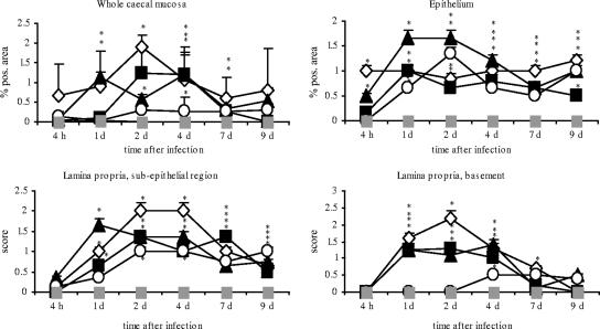
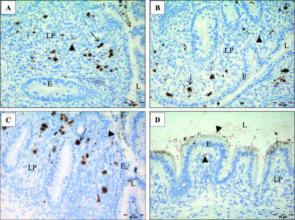
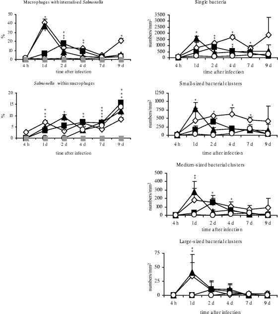
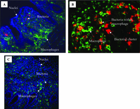
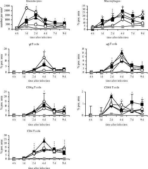
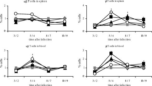
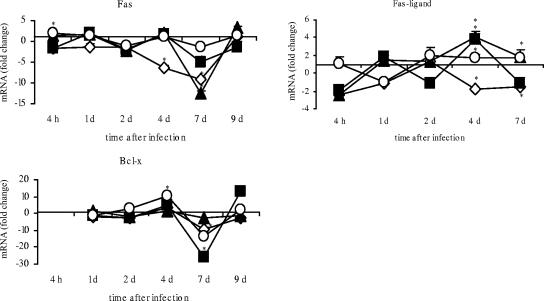
Day-old chicks are very susceptible to infections with Salmonella enterica subspecies. The gut mucosa is the initial site of host invasion and provides the first line of defense against the bacteria. To study the potential of different S. enterica serovars to invade the gut mucosa and trigger an immune response, day-old chicks were infected orally with Salmonella enterica serovar Enteritidis, S. enterica serovar Typhimurium, S. enterica serovar Hadar, or S. enterica serovar Infantis, respectively. The localization of Salmonella organisms in gut mucosa and the number of immune cells in cecum were determined by immunohistochemistry in the period between 4 h and 9 days after infection. Using quantitative real-time reverse transcription (RT)-PCR, mRNA expression of various cytokines, chemokines, and inducible nitric oxide synthase (iNOS) was examined in cecum. As a result, all S. enterica serovars were able to infect epithelial cells and the lamina propria. Notably, serovar Enteritidis showed the highest invasiveness of lamina propria tissue, whereas serovars Typhimurium and Hadar displayed moderate invasiveness and serovar Infantis hardly any invasion capabilities. Only a limited number of bacteria of all serovars were found within intestinal macrophages. Elevated numbers of granulocytes, CD8+ cells, and TCR1+ cells and mRNA expression rates for interleukin 12 (IL-12), IL-18, tumor necrosis factor alpha factor, and iNOS in cecum correlated well with the invasiveness of serovars in the lamina propria. In contrast, changes in numbers of TCR2+ and CD4+ cells and IL-2 mRNA expression seemed to be more dependent on infection of epithelial cells. The data indicate that the capability of Salmonella serovars to enter the cecal mucosa and invade lower regions affects both the level and character of the immune response in tissue.


Figures

FIG. 1.
FIG. 1.
Diagrams showing percentages of Salmonella positive-stained areas in whole avian cecum mucosa and epithelium and the scores for numbers of bacteria in subepithelial and basement regions of cecum lamina propria after infection with different Salmonella serovars. Asterisks indicate a significant difference between the treated and control groups (P ≤ 0.05). ⋄, serovar Enteritidis; ▪, serovar Typhimurium; ▴, serovar Hadar; ○, serovar Infantis; ░⃞, control. d, day; pos., positive.
FIG. 2.
FIG. 2.
Representative photographs of cecum mucosa after infection of day-old chicks with different Salmonella serovars. Single bacteria are marked with arrowheads and bacterial accumulations with arrows. (A) Serovar Enteritidis infection, 2 dpi: bacteria spread over the whole lamina propria. (B) Serovar Typhimurium infection at 2 dpi: bacteria predominantly located subepithelially. (C) Serovar Hadar infection at 2 dpi: bacteria predominantly located subepithelially. (D) Serovar Infantis infection at 2 dpi: bacteria are predominantly located in epithelial lining.
FIG. 3.
FIG. 3.
Percentages of Salmonella organisms within CVI 68.1-stained macrophages and Salmonella-positive macrophages and numbers of single bacteria and variably sized bacterial clusters per mm2 after infection of day-old chicks with different Salmonella serovars in cecum mucosa. Asterisks indicate a significant difference between the treated and control groups (P ≤ 0.05). ⋄, serovar Enteritidis; ▪, serovar Typhimurium; ▴, serovar Hadar; ○, serovar Infantis; ░⃞, control. d, day.
FIG. 4.
FIG. 4.
Representative confocal laser-scanning micrographs of cecum mucosa of serovar Typhimurium-infected (A) or serovar Enteritidis-infected (B and C) chicks. Red, Salmonella; green, CVI 68.1-positive macrophages; blue, cell nuclei. (A) 1 dpi; different localizations of bacteria (mainly subepithelial) and macrophages (mainly in basement regions) are shown at this time point after infection. Magnification, ×20. (B) 1 dpi; representative region showing colocalization of macrophages and bacteria and Salmonella clusters outside macrophages. Magnification, ×40. (C) 4 dpi; many single bacteria distributed over the whole lamina propria and not colocalized with macrophages. Magnification, ×20.
FIG. 5.
FIG. 5.
Occurrence of immune cells after oral infection of day-old chicks with different Salmonella serovars in cecum. Asterisks indicate a significant difference between the treated and control groups (P ≤ 0.05). ⋄, serovar Enteritidis; ▪, serovar Typhimurium; ▴, serovar Hadar; ○, serovar Infantis; ░⃞, control. d, day; pos., positive.
FIG. 6.
FIG. 6.
Relative proportions of alpha/beta and gamma/delta T cells in blood and spleen. Day-old chicks were orally infected with different Salmonella serovars. Results for control animals were set to 1. Asterisks indicate a significant difference between the treated and control groups (P ≤ 0.05). ⋄, serovar Enteritidis; ▪, serovar Typhimurium; ▴, serovar Hadar; ○, serovar Infantis; ░⃞, control. The x axis for each panel shows days postinfection.
FIG. 7.
FIG. 7.
Diagrams showing quantifications of different cytokines, chemokines, and iNOS after infection of day-old chicks with different Salmonella serovars. Data are given as n-fold changes in mRNA levels for infected birds relative to levels for noninfected controls. Each value represents the mean for three individual samples. Standard errors are shown as vertical bars. Very small standard deviations are often not visible. Asterisks indicate a significant difference between the treated and control groups (P ≤ 0.05). ⋄, serovar Enteritidis; ▪, serovar Typhimurium; ▴, serovar Hadar; ○, serovar Infantis. d, day.
FIG. 8.
FIG. 8.
Quantification of Fas, FasL, and Bcl-x expression after infection of day-old chicks with different Salmonella serovars. Data are given as n-fold changes in mRNA levels for infected birds relative to those for noninfected controls. Each value represents the mean for three individual samples. Standard errors are shown as vertical bars. The very small standard deviations are often not visible. Asterisks indicate a significant difference between the treated and control groups (P ≤ 0.05). ⋄, serovar Enteritidis; ▪, serovar Typhimurium; ▴, serovar Hadar; ○, serovar Infantis. d, day.

References

    1. Aabo, S., J. P. Christensen, M. S. Chadfield, B. Carstensen, J. E. Olsen, and M. Bisgaard. 2002. Quantitative comparison of intestinal invasion of zoonotic serotypes of Salmonella enterica in poultry. Avian Pathol. 31:41-47. - PubMed
    1. Asheg, A., M. Levkut, V. Revajova, Z. Sevcikova, L. Kolodzieyski, and J. Pistl. 2002. T lymphocyte subpopulations and B lymphocyte cells in caecum and spleen of chicks infected with Salmonella enteritidis. Acta Histochem. 104:435-439. - PubMed
    1. Babu, U., M. Scott, M. J. Myers, M. Okamura, D. Gaines, H. F. Yancy, H. Lillehoj, R. A. Heckert, and R. B. Raybourne. 2003. Effects of live attenuated and killed Salmonella vaccine on T-lymphocyte mediated immunity in laying hens. Vet. Immunol. Immunopathol. 91:39-44. - PubMed
    1. Beal, R. K., C. Powers, T. F. Davison, P. A. Barrow, and A. L. Smith. 2006. Clearance of enteric Salmonella enterica serovar Typhimurium in chickens is independent of B-cell function. Infect. Immun. 74:1442-1444. - PMC - PubMed
    1. Beal, R. K., C. Powers, P. Wigley, P. A. Barrow, P. Kaiser, and A. L. Smith. 2005. A strong antigen-specific T-cell response is associated with age and genetically dependent resistance to avian enteric salmonellosis. Infect. Immun. 73:7509-7516. - PMC - PubMed

Publication types

MeSH terms