. 2007 Nov;75(11):5465-75.
doi: 10.1128/IAI.00822-07. Epub 2007 Aug 20.

L-fucose stimulates utilization of D-ribose by Escherichia coli MG1655 DeltafucAO and E. coli Nissle 1917 DeltafucAO mutants in the mouse intestine and in M9 minimal medium

Affiliations

L-fucose stimulates utilization of D-ribose by Escherichia coli MG1655 DeltafucAO and E. coli Nissle 1917 DeltafucAO mutants in the mouse intestine and in M9 minimal medium

Steven M Autieri et al. Infect Immun. 2007 Nov.

Abstract

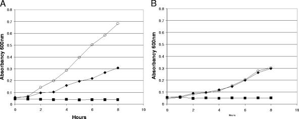
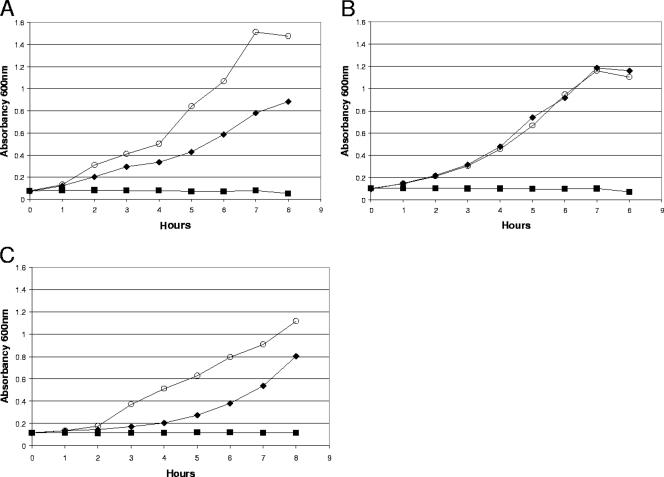
Escherichia coli MG1655 uses several sugars for growth in the mouse intestine. To determine the roles of L-fucose and D-ribose, an E. coli MG1655 DeltafucAO mutant and an E. coli MG1655 DeltarbsK mutant were fed separately to mice along with wild-type E. coli MG1655. The E. coli MG1655 DeltafucAO mutant colonized the intestine at a level 2 orders of magnitude lower than that of the wild type, but the E. coli MG1655 DeltarbsK mutant and the wild type colonized at nearly identical levels. Surprisingly, an E. coli MG1655 DeltafucAO DeltarbsK mutant was eliminated from the intestine by either wild-type E. coli MG1655 or E. coli MG1655 DeltafucAO, suggesting that the DeltafucAO mutant switches to ribose in vivo. Indeed, in vitro growth experiments showed that L-fucose stimulated utilization of D-ribose by the E. coli MG1655 DeltafucAO mutant but not by an E. coli MG1655 DeltafucK mutant. Since the DeltafucK mutant cannot convert L-fuculose to L-fuculose-1-phosphate, whereas the DeltafucAO mutant accumulates L-fuculose-1-phosphate, the data suggest that L-fuculose-1-phosphate stimulates growth on ribose both in the intestine and in vitro. An E. coli Nissle 1917 DeltafucAO mutant, derived from a human probiotic commensal strain, acted in a manner identical to that of E. coli MG1655 DeltafucAO in vivo and in vitro. Furthermore, L-fucose at a concentration too low to support growth stimulated the utilization of ribose by the wild-type E. coli strains in vitro. Collectively, the data suggest that L-fuculose-1-phosphate plays a role in the regulation of ribose usage as a carbon source by E. coli MG1655 and E. coli Nissle 1917 in the mouse intestine.


Figures

FIG. 1.
FIG. 1.
Fucose operons and degradation pathway (A) and the ribose operon and degradation pathway (B). Arrows above genes indicate promoters and the direction of transcription. MFS, major facilitator superfamily; ABC, ATP-binding cassette.
FIG. 2.
FIG. 2.
Colonization of the mouse intestine by E. coli MG1655 ΔfucAO, E. coli MG1655 ΔrbsK, and E. coli MG1655 ΔfucAO ΔrbsK. Sets of three mice were fed 105 CFU of E. coli MG1655 (▪) and 105 CFU of MG1655 ΔfucAO (○) (A); 105 CFU of E. coli MG1655 (▪) and 105 CFU of E. coli MG1655 ΔrbsK (○) (B); 105 CFU of E. coli MG1655 (▪) and 105 CFU of E. coli MG1655 ΔfucAO ΔrbsK (○) (C); 105 CFU of E. coli MG1655 ΔfucAO (▪) and 105 CFU of E. coli MG1655 ΔfucAO ΔrbsK (○) (D); and 1010 CFU of E. coli MG1655 (▪) and 105 CFU of E. coli MG1655 ΔfucAO (○) (E). At the indicated times, fecal samples were homogenized, diluted, and plated as described in Materials and Methods. Bars representing standard errors of the log10 means of CFU per gram of feces for six mice are presented for each time point except for panels A and B, for which data from 12 mice are presented. Each colonization curve has the specific strain genotype immediately above or below it. wt, wild-type E. coli MG1655.
FIG. 3.
FIG. 3.
Colonization of the mouse intestine by E. coli MG1655 ΔfucK. Sets of three mice were fed 105 CFU of E. coli MG1655 (▪) and 105 CFU of MG1655 ΔfucK (○) (A) or 1010 CFU of E. coli MG1655 (▪) and 105 CFU of E. coli MG1655 ΔfucK (○) (B). At the indicated times, fecal samples were homogenized, diluted, and plated as described in Materials and Methods. Bars representing standard errors of the log10 means of CFU per gram of feces for 12 mice are presented for each time point in panel A and for 6 mice in panel B. Each colonization curve has the specific strain genotype immediately above or below it. wt, wild-type E. coli MG1655.
FIG. 4.
FIG. 4.
Growth of E. coli MG1655 ΔfucAO (A) and E. coli MG1655 ΔfucK (B) in M9 minimal medium in the presence of 0.05% (wt/wt) fucose (▪), 0.15% (wt/wt) ribose (⧫), or a mixture of 0.05% (wt/wt) fucose and 0.15% (wt/wt) ribose (○). E. coli MG1655 ΔfucAO and E. coli MG1655 ΔfucK were grown in M9 minimal medium containing glycerol (0.2%, wt/wt), washed, and resuspended in M9 minimal medium containing the appropriate sugars (see Materials and Methods). Incubation was at 37°C with aeration. A600 readings at the indicated times are presented. Growth experiments were performed at least three times. The results of typical experiments are shown.
FIG. 5.
FIG. 5.
Growth of E. coli MG1655 ΔfucAO in M9 minimal medium in the presence of 0.05% (wt/wt) fucose (▪), 0.15% (wt/wt) N-acetylglucosamine (⧫), or a mixture of 0.05% (wt/wt) fucose and 0.15% (wt/wt) N-acetylglucosamine (○) (A); 0.05% (wt/wt) fucose (▪), 0.15% (wt/wt) mannose (⧫), or a mixture of 0.05% (wt/wt) fucose and 0.15% (wt/wt) mannose (○) (B); and 0.05% (wt/wt) fucose (▪), 0.15% (wt/wt) galactose (⧫), or a mixture of 0.05% (wt/wt) fucose and 0.15% (wt/wt) galactose (○) (C). E. coli MG1655 ΔfucAO was grown in M9 minimal medium containing glycerol (0.2%, wt/wt), washed, and resuspended in M9 minimal medium containing the appropriate sugars (see Materials and Methods). Incubation was at 37°C with aeration. A600 readings at the indicated times are presented. Growth experiments were performed at least three times. The results of typical experiments are shown.
FIG. 6.
FIG. 6.
Growth of wild-type E. coli MG1655 in M9 minimal medium in the presence of 0.005% (wt/wt) fucose (▪), 0.15% (wt/wt) ribose (⧫), or a mixture of 0.005% (wt/wt) fucose and 0.15% (wt/wt) ribose (○). Wild-type E. coli MG1655 was grown in M9 minimal medium containing glycerol (0.2%, wt/wt), washed, and resuspended in M9 minimal medium containing the appropriate sugars (see Materials and Methods). Incubation was at 37°C with aeration. A600 readings at the indicated times are presented. Growth experiments were performed at least three times. The results of typical experiments are shown.
FIG. 7.
FIG. 7.
Growth of E. coli Nissle 1917 ΔfucAO in M9 minimal medium in the presence of 0.05% (wt/wt) fucose (▪), 0.15% (wt/wt) ribose (⧫), or a mixture of 0.05% (wt/wt) fucose and 0.15% (wt/wt) ribose (○) (A); growth of E. coli Nissle 1917 ΔfucK in M9 minimal medium in the presence of 0.05% (wt/wt) fucose (▪), 0.15% (wt/wt) ribose (⧫), or a mixture of 0.05% (wt/wt) fucose and 0.15% (wt/wt) ribose (○) (B); and growth of wild-type E. coli Nissle 1917 in M9 minimal medium in the presence of 0.005% (wt/wt) fucose (▪), 0.15% (wt/wt) ribose (⧫), or a mixture of 0.005% (wt/wt) fucose and 0.15% (wt/wt) ribose (○) (C). Strains were grown in M9 minimal medium containing glycerol (0.2%, wt/wt), washed, and resuspended in M9 minimal medium containing the appropriate sugars (see Materials and Methods). Incubation was at 37°C with aeration. A600 readings at the indicated times are presented. Growth experiments were performed at least three times. The results of typical experiments are shown.

References

    1. Allan, A. 1981. Structure and function of gastrointestinal mucus, p. 637-639. In L. R. Johnson (ed.), Physiology of the gastrointestinal tract. Raven Press, New York, NY.
    1. Anderson, J. D., W. A. Gillespie, and M. H. Richmond. 1973. Chemotherapy and antibiotic-resistance transfer between enterobacteria in the human gastro-intestinal tract. J. Med. Microbiol. 6:461-473. - PubMed
    1. Atuma, C., V. Strugala, A. Allen, and L. Holm. 2001. The adherent gastrointestinal mucus gel layer: thickness and physical state in vivo. Am. J. Physiol. Gastrointest. Liver Physiol. 280:G922-G929. - PubMed
    1. Bartkus, J. M., and R. P. Mortlock. 1986. Isolation of a mutation resulting in constitutive synthesis of l-fucose catabolic enzymes. J. Bacteriol. 165:710-714. - PMC - PubMed
    1. Blattner, F. R., G. Plunkett III, C. A. Bloch, N. T. Perna, V. Burland, M. Riley, J. Collado-Vides, J. D. Glasner, C. K. Rode, G. F. Mayhew, J. Gregor, N. W. Davis, H. A. Kirkpatrick, M. A. Goeden, D. J. Rose, B. Mau, and Y. Shao. 1997. The complete genome sequence of Escherichia coli K-12. Science 277:1453-1474. - PubMed

Publication types

LinkOut - more resources