

doi: 10.4049/jimmunol.179.5.2961.
Airway hyperresponsiveness through synergy of gammadelta} T cells and NKT cells
Affiliations
- PMID: 17709511
- PMCID: PMC4480876
- DOI: 10.4049/jimmunol.179.5.2961
Item in Clipboard
Airway hyperresponsiveness through synergy of gammadelta} T cells and NKT cells
J Immunol.
.
Abstract
Mice sensitized and challenged with OVA were used to investigate the role of innate T cells in the development of allergic airway hyperresponsiveness (AHR). AHR, but not eosinophilic airway inflammation, was induced in T cell-deficient mice by small numbers of cotransferred gammadelta T cells and invariant NKT cells, whereas either cell type alone was not effective. Only Vgamma1+Vdelta5+ gammadelta T cells enhanced AHR. Surprisingly, OVA-specific alphabeta T cells were not required, revealing a pathway of AHR development mediated entirely by innate T cells. The data suggest that lymphocytic synergism, which is key to the Ag-specific adaptive immune response, is also intrinsic to T cell-dependent innate responses.
Figures
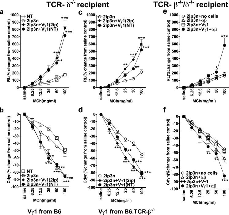
Vγ1+
γδ T cells from naive donors enhance AHR when αβ T cells are present. AHR was monitored by measuring RL (a, c, and e) and Cdyn (b, d, and f). a and b, Reconstitution of γδ T cell-deficient mice with Vγ1+ cells restores AHR. OVA-sensitized B6.TCR-δ−/− mice received 1 × 10⁴ splenic Vγ1+
γδ T cells from untreated (NT) or sensitized (2ip) C57BL/6 donors, before airway challenge. Untreated recipients (NT) and recipients that were sensitized and challenged, but did not receive cells (2ip3N), are also shown. Results for each group are presented as means ± SEM (n = 8). Significant differences between 2ip3N and 2ip3N + Vγ1 groups are indicated: **, p < 0.01; ***, p < 0.001. c and d, Vγ1+ cells from αβ T cell-deficient mice are still capable of restoring AHR. OVA-sensitized B6.TCR-δ−/− mice received 1 × 10⁴ splenic Vγ1+
γδ T cells from untreated (NT) or sensitized (2ip) B6.TCR-β−/− donors, before airway challenge. Recipients that were sensitized and challenged, but did not receive cells (2ip3N), are also shown. Results for each group are presented as means ± SEM (n = 12). Significant differences between 2ip3N and 2ip3N + Vγ1 groups are indicated as follows: *, p < 0.05; **, p < 0.01; ***, p < 0.001. e and f, Vγ1+ cells together with αβ T cells restore AHR in T cell-deficient mice. OVA-sensitized B6.TCR-β−/−δ−/− mice received 1 × 10⁴ splenic Vγ1+
γδ T cells from sensitized B6.TCR-β−/− donors and 2 × 10⁶
αβ T cells from sensitized B6.TCR-δ−/− donors (Vγ1 + αβ), before airway challenge. Recipients that were sensitized and challenged, but did not receive cells (2ip3N), or received only Vγ1+ cells (Vγ1) or only αβ T cells (αβ), are also shown. Results for each group are presented as means ± SEM (n = 4–9). Significant differences between sensitized and challenged mice, which received no cells, or Vγ1+ cells plus αβ T cells, are indicated as follows: *, p < 0.05; **, p < 0.01; ***, p < 0.001.
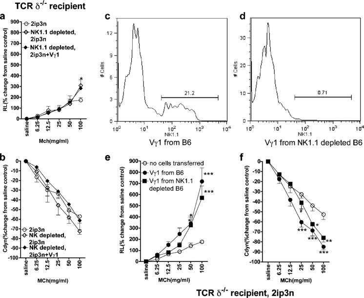
Vγ1+
γδ T cells fail to mediate AHR when NK1.1+ cells are absent. AHR was monitored by measuring RL (a and e) and Cdyn (b and f). a and b, Vγ1+ cells fail to restore AHR in γδ T cell-deficient mice pretreated with anti-NK1.1 mAb. OVA-sensitized B6.TCR-δ−/− mice were treated with mAb PK136 (200 μg i.v.), received 1 × 10⁴ splenic Vγ1+
γδ T cells 3 days later, and were then challenged via the airways (NK1.1-depleted, 2ip3N + Vγ1). Recipients that were only sensitized and challenged (2ip3N) and those that were sensitized and challenged and treated with the Ab (NK1.1-depleted, 2ip3N) are also shown. Results for each group are presented as means ± SEM (n = 4). Significant differences between 2ip3N and 2ip3N, NK1.1-depleted groups are indicated as follows: *, p < 0.05. c and d, Depletion of NK1.1+ cells within the Vγ1+ population. C57BL/6 mice were treated with mAb PK136 (200 μg i.v.) and 3 days later, NAD splenocytes were stained for TCR-δ, Vγ1, and NK1.1. Cytofluorimetric analysis shows that ~20% of gated splenic Vγ1+
γδ T cells express NK1.1 (c) and that the treatment with mAb PK136 removes most of these cells (d). e and f, NK1.1− Vγ1+
γδ T cells enhance AHR. OVA-sensitized B6.TCR-δ−/− mice received 1 × 10⁴ splenic Vγ1+
γδ T cells from C57BL/6 donors before airway challenge. The cell donors were either untreated or received mAb PK136 i.v., 3 days before cell transfer (Vγ1 from B6 and Vγ1 from NK1.1-depleted B6). Recipients that were sensitized and challenged, but did not receive cells, are also shown (no cells transferred). Results for each group are presented as means ± SEM (n = 7–8). Significant differences between mice that received no cells or Vγ1 cells are indicated as follows: *, p < 0.05; **, p < 0.01; ***, p < 0.001. Significant difference between mice that had received Vγ1+ cells from B6 vs from NK1.1-depleted B6 (f) is as follows: #, p < 0.05.
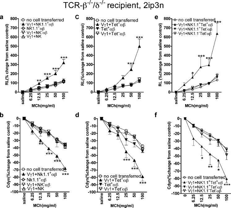
Vγ1+
γδ T cells synergize with NKT cells in mediating AHR. AHR was monitored by measuring RL (a, c, and e) and Cdyn (b, d, and f). a and b, Vγ1+ cells together with NK1.1+
αβ T cells restore AHR in T cell-deficient mice. OVA-sensitized B6.TCR-β−/−δ−/− mice received 1 × 10⁴ splenic Vγ1+
γδ T cells from sensitized B6.TCR-β−/− donors and 2 × 10⁴ NK1.1+
αβ T cells from sensitized B6.TCR-δ−/− donors (Vγ1 + NK1.1+αβ), before airway challenge. Recipients that were sensitized and challenged, but did not receive cells (no cell transferred), or that received 2 × 10⁴ NK1.1+
αβ T cells (NK1.1+αβ) alone, Vγ1+ cells plus 2 × 10⁶ NK1.1−
αβ T cells (Vγ1 + NK1.1−αβ), or Vγ1+ cells plus 9 × 10⁴ NK1.1+ non-T cells (Vγ1 + NK), are also shown. Results for each group are presented as means ± SEM (n = 6–7). Significant differences between mice that received no cells or Vγ1+ cells plus NK1.1+
αβ T cells are indicated as follows: **, p < 0.01; ***, p < 0.001. c and d, Vγ1+ cells together with CD1d tetramer+
αβ T cells restore AHR in T cell-deficient mice. OVA-sensitized B6.TCR-β−/−δ−/− mice received 1 × 10⁴ splenic Vγ1+
γδ T cells from sensitized B6.TCR-β−/− donors and 2 × 10⁴ CD1d tetramer+
αβ T cells from sensitized B6.TCR-δ−/− donors (Vγ1 + Tet+
αβ), before airway challenge. Recipients that were sensitized and challenged, but did not receive cells (no cells), or that received 2 × 10⁴ Tet+
αβ T cells (Tet+
αβ) alone, or Vγ1+ cells plus 2 × 10⁴ Tet- αβ T cells (Vγ1 + Tet−
αβ), are also shown. Results for each group are presented as means ± SEM (n = 4–5). Significant differences between mice that received no cells or Vγ1+ plus NK1.1+
αβ T cells are indicated as follows: ***, p < 0.001. e and f, Only NK1.1+
αβ T cells that are also CD1d tetramer+ synergize with Vγ1+
γδ T cells in mediating AHR. OVA-sensitized B6.TCR-β−/−δ−/− mice received 1 × 10⁴ splenic Vγ1+
γδ T cells from sensitized B6.TCR-β−/− mice and 2 × 10⁴ NK1.1+ CD1d tetramer+
αβ T cells from sensitized B6.TCR-δ−/− donors (Vγ1 + NK1.1+Tet+
αβ), before airway challenge. Recipients that were sensitized and challenged, but did not receive cells (no cell transferred), or that received Vγ1+ cells and 2 × 10⁴ NK1.1+Tet−
αβ T cells (Vγ1 + NK1.1+Tet−
αβ), or Vγ1+ cells plus 2 × 10⁴ NK1.1−Tet−
αβ T cells (Vγ1 + NK1.1−Tet−
αβ), are also shown. Results for each group are presented as means ± SEM (n = 5–6). Significant differences between mice that received no cells or Vγ1+ cells plus NK1.1+
αβ T cells are indicated as follows: *, p < 0.05; **, p < 0.01; ***, p < 0.001.
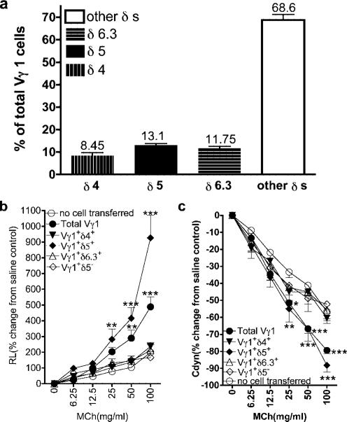
Comparison of Vγ1+
γδ T cells expressing different Vδs for their ability to mediate AHR. a, Vδ expression among Vγ1+
γδ T cells in B6.TCR-β−/− spleen. NAD splenocytes of adult B6.TCR-β−/− mice were stained with Abs against Vγ1, TCR-δ, and several Vδs, and analyzed cytofluorimetrically. Results for each group are presented as means ± SEM (n = 5–11). b and c, Reconstitution of γδ T cell-deficient mice with Vδ+ fractions of Vγ1+ cells. OVA-sensitized B6.TCR-δ−/− mice received 1 × 10⁴ sorted Vγ1+
γδ T cells from untreated B6.TCR-β−/− spleen, expressing the indicated Vδs, before airway challenge (2ip3N + Vγ1). Recipients that were sensitized and challenged, but did not receive cells (no cell transferred), are also shown. AHR was monitored by measuring RL (b) and Cdyn (c). Results for each group are presented as means ± SEM (n = 5–8). Significant differences between mice that received no cells or Vγ1+ cells are indicated as follows: **, p < 0.01; ***, p < 0.001.
Similar articles
-
Allergic airway hyperresponsiveness-enhancing gammadelta T cells develop in normal untreated mice and fail to produce IL-4/13, unlike Th2 and NKT cells.J Immunol. 2009 Feb 15;182(4):2002-10. doi: 10.4049/jimmunol.0803280. J Immunol. 2009. PMID: 19201853 Free PMC article.
-
Vgamma1+ T cells and tumor necrosis factor-alpha in ozone-induced airway hyperresponsiveness.Am J Respir Cell Mol Biol. 2009 Apr;40(4):454-63. doi: 10.1165/rcmb.2008-0346OC. Epub 2008 Oct 16. Am J Respir Cell Mol Biol. 2009. PMID: 18927346 Free PMC article.
-
gammadelta T cells contribute to the systemic immunoglobulin E response and local B-cell reactivity in allergic eosinophilic airway inflammation.Immunology. 2003 Jan;108(1):98-108. doi: 10.1046/j.1365-2567.2003.01561.x. Immunology. 2003. PMID: 12519308 Free PMC article.
-
Role of IgE in the development of allergic airway inflammation and airway hyperresponsiveness--a murine model.Allergy. 1999 Apr;54(4):297-305. doi: 10.1034/j.1398-9995.1999.00085.x. Allergy. 1999. PMID: 10371087 Review.
-
Anti-tumour immunotherapy with Vγ9Vδ2 T lymphocytes: from the bench to the bedside.Br J Haematol. 2013 Jan;160(2):123-32. doi: 10.1111/bjh.12090. Epub 2012 Oct 15. Br J Haematol. 2013. PMID: 23061882 Review.
Cited by
-
ICOS/ICOSL interaction is required for CD4+ invariant NKT cell function and homeostatic survival.J Immunol. 2008 Apr 15;180(8):5448-56. doi: 10.4049/jimmunol.180.8.5448. J Immunol. 2008. PMID: 18390727 Free PMC article.
-
Alternative spliced CD1d transcripts in human bronchial epithelial cells.PLoS One. 2011;6(8):e22726. doi: 10.1371/journal.pone.0022726. Epub 2011 Aug 11. PLoS One. 2011. PMID: 21853044 Free PMC article.
-
Aligning mouse models of asthma to human endotypes of disease.Respirology. 2014 Aug;19(6):823-33. doi: 10.1111/resp.12315. Epub 2014 May 9. Respirology. 2014. PMID: 24811131 Free PMC article. Review.
-
A novel role for NKT cells in cutaneous wound repair.J Surg Res. 2011 Jun 15;168(2):325-33.e1. doi: 10.1016/j.jss.2009.09.030. Epub 2009 Oct 9. J Surg Res. 2011. PMID: 20089261 Free PMC article.
-
Evidence that CD8+ dendritic cells enable the development of gammadelta T cells that modulate airway hyperresponsiveness.J Immunol. 2008 Jul 1;181(1):309-19. doi: 10.4049/jimmunol.181.1.309. J Immunol. 2008. PMID: 18566396 Free PMC article.
References
-
- Claman HN, Chaperon EA, Triplett RF. Immunocompetence of transferred thymus-marrow cell combinations. J. Immunol. 1966;97:828–832. - PubMed
-
- Castellino F, Huang AY, Altan-Bonnet G, Stoll S, Scheinecker C, Germain RN. Chemokines enhance immunity by guiding naive CD8+ T cells to sites of CD4+ T cell-dendritic cell interaction. Nature. 2006;440:890–895. - PubMed
-
- Wills-Karp M. Immunologic basis of antigen-induced airway hyperresponsiveness. Annu. Rev. Immunol. 1999;17:255–281. - PubMed
-
- Robinson DS, Hamid O, Ying S, Tsicopoulos A, Barkans J, Bentley AM, Corrigan C, Durham SR, Kay AB. Predominant TH2-like bronchoalveolar T-lymphocyte population in atopic asthma. N. Engl. J. Med. 1992;326:298–304. - PubMed
Publication types
MeSH terms
Substances
Grants and funding
- P01 HL036577/HL/NHLBI NIH HHS/United States
- R01 AI044920/AI/NIAID NIH HHS/United States
- HL 61005/HL/NHLBI NIH HHS/United States
- HL 36577/HL/NHLBI NIH HHS/United States
- AI 44920/AI/NIAID NIH HHS/United States
- HL 65410/HL/NHLBI NIH HHS/United States
- R21 AI063400/AI/NIAID NIH HHS/United States
- AI 40611/AI/NIAID NIH HHS/United States
- R01 HL061005/HL/NHLBI NIH HHS/United States
- R01 HL065410/HL/NHLBI NIH HHS/United States
- AI 063400/AI/NIAID NIH HHS/United States
- R01 AI057485/AI/NIAID NIH HHS/United States
- R01 AI040611/AI/NIAID NIH HHS/United States
- AI 057485/AI/NIAID NIH HHS/United States
LinkOut - more resources
Full Text Sources
Other Literature Sources
