. 2007 Aug 28;104(35):14068-73.
doi: 10.1073/pnas.0706760104. Epub 2007 Aug 20.

Human cardiac stem cells

Affiliations

Human cardiac stem cells

Claudia Bearzi et al. Proc Natl Acad Sci U S A. .

Abstract

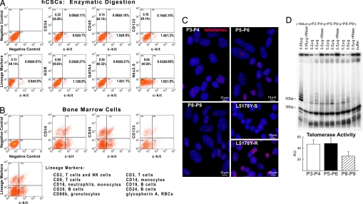
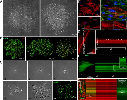
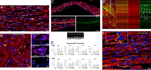
The identification of cardiac progenitor cells in mammals raises the possibility that the human heart contains a population of stem cells capable of generating cardiomyocytes and coronary vessels. The characterization of human cardiac stem cells (hCSCs) would have important clinical implications for the management of the failing heart. We have established the conditions for the isolation and expansion of c-kit-positive hCSCs from small samples of myocardium. Additionally, we have tested whether these cells have the ability to form functionally competent human myocardium after infarction in immunocompromised animals. Here, we report the identification in vitro of a class of human c-kit-positive cardiac cells that possess the fundamental properties of stem cells: they are self-renewing, clonogenic, and multipotent. hCSCs differentiate predominantly into cardiomyocytes and, to a lesser extent, into smooth muscle cells and endothelial cells. When locally injected in the infarcted myocardium of immunodeficient mice and immunosuppressed rats, hCSCs generate a chimeric heart, which contains human myocardium composed of myocytes, coronary resistance arterioles, and capillaries. The human myocardium is structurally and functionally integrated with the rodent myocardium and contributes to the performance of the infarcted heart. Differentiated human cardiac cells possess only one set of human sex chromosomes excluding cell fusion. The lack of cell fusion was confirmed by the Cre-lox strategy. Thus, hCSCs can be isolated and expanded in vitro for subsequent autologous regeneration of dead myocardium in patients affected by heart failure of ischemic and nonischemic origin.


Conflict of interest statement

Conflict of interest statement: P.A. has applied for a patent.

Figures

Fig. 1.
Fig. 1.
Cardiac niches and hCSC division. Sections of normal human myocardium. (A–C) Cluster of c-kitPOS cells (green). Arrows in A define the areas in B and C. Gap (connexin 43: Cx43, white; arrowheads) and adherens (N-cadherin: N-cadh, magenta; arrowheads) junctions are shown at higher magnification. Cx43 and N-cadh are present between c-kitPOS cells and myocytes (α-SA, red) and fibroblasts (procollagen, light blue); fibronectin, yellow. (D and E) Mitosis (phospho-H3, magenta; arrows) in c-kitPOS cells; α-adaptin (D, white) and Numb (E, yellow) show a uniform (D) and nonuniform (E) localization in the mitotic c-kitPOS cells.
Fig. 2.
Fig. 2.
Human CSCs. (A and B) Scatter plots of hCSCs (A) and human bone marrow cells (B). hCSCs do not express hematopoietic markers, KDR, GATA4, and Nkx2.5. (C) Nuclei (blue) of hCSCs were stained with a telomere probe (magenta). Lymphoma cells with short (7-kbp) and long (48-kbp) telomeres are shown for comparison. (D) Products of telomerase activity in hCSCs start at 50 bp and display a 6-bp periodicity. Samples treated with RNase and CHAPS buffer were used as negative controls, and HeLa cells were used as positive control. The band at 36 bp corresponds to an internal control for PCR efficiency. Optical density (arbitrary units, AU) is shown as mean ± SD.
Fig. 3.
Fig. 3.
In vitro properties of hCSCs. (A–C) Clones formed by hCSCs (c-kit, green) isolated by enzymatic digestion (A and C) or primary explant (B). The number of cells increased with time (C). (D) hCSCs generate myocytes positive for cardiac myosin heavy-chain (MHC), α-SA, and α-cardiac-actinin (α-actinin). Sarcomeres are apparent (Insets); phalloidin, green. (E) Myocyte shortening in cells derived from clonogenic hCSCs was recorded by two-photon microscopy and laser line-scan imaging (Left). The line scan is shown (Right), and arrowheads point to individual contractions. (F) Myocytes derived from EGFP-positive hCSCs, cocultured with neonatal myocytes. EGFP-positive human myocytes shorten (arrowheads) with electrical stimulation. (G) Calcium transients in EGFP-positive human myocytes and EGFP-negative rat myocytes (calcium indicator Rhod-2, red).
Fig. 4.
Fig. 4.
hCSCs regenerate infarcted myocardium. (A) Mouse heart 21 days after infarction and injection of hCSCs. Human myocardium (arrowheads) is present within the infarct (MI). BZ, border zone. Areas in rectangles are shown at higher magnification below. Human myocytes are α-SA- (red) and Alu- (green) positive. Asterisks indicate spared myocytes. (B) Expression of human (h) genes by real-time RT-PCR in treated infarcted rats at 5–11 and 12–21 days. Clonogenic hCSCs were used for comparison of human transcripts. (C) Electrophoresis of real-time RT-PCR products (for sequences see SI Fig. 11J).
Fig. 5.
Fig. 5.
Integration of human myocardium. (A) Human myocytes are Cre-recombinase-positive (white) but EGFP negative. (B) Human myocytes and vessels show, at most, two human X-chromosomes (X-Chr, white dots; arrowheads). Mouse X-Chr (magenta dots; arrows) are present in myocytes of the border zone (BZ). (C) Transmural infarct in a treated rat; human myocardium (arrowheads) is present within the infarct. The area in the rectangle is shown at higher magnification (Bottom); human myocytes are α-SA- (red) and Alu- (green) positive. Echocardiogram shows contraction in the infarcted wall (arrowheads). (D) Ventricular function. Results are mean ± SD. * and †, Difference, P < 0.05, versus SO (sham-operated) and MI, respectively. (E) Calcium transient in EGFP-positive human myocytes and EGFP-negative mouse myocytes recorded by two-photon microscopy and laser line-scan imaging (calcium indicator Rhod-2, red). (F) Myocardial regeneration at 3 weeks. Connexin 43 (Cx43, yellow) is present between human-myocytes (α-SA, red; Alu, green) and spared rat myocytes (α-SA, red; Alu-negative). See Inset for higher magnification.

Similar articles

Cited by

References

    1. Leri A, Kajstura J, Anversa P. Physiol Rev. 2005;85:1373–1416. - PubMed
    1. Messina E, De Angelis L, Frati G, Morrone S, Chimenti S, Fiordaliso F, Salio M, Battaglia M, Latronico MVG, Coletta M, et al. Circ Res. 2004;95:911–921. - PubMed
    1. Urbanek K, Torella D, Sheikh F, De Angelis A, Nurzynska D, Silvestri F, Beltrami CA, Bussani R, Beltrami AP, Quaini F, et al. Proc Natl Acad Sci USA. 2005;102:8692–8697. - PMC - PubMed
    1. Beltrami AP, Barlucchi L, Torella D, Baker M, Limana F, Chimenti S, Kasahara H, Rota M, Musso E, Urbanek K, et al. Cell. 2003;114:763–766. - PubMed
    1. Linke A, Muller P, Nurzynska D, Casarsa C, Torella D, Nascimbene A, Castaldo C, Cascapera S, Bohm M, Quaini F, et al. Proc Natl Acad Sci USA. 2005;102:8966–8971. - PMC - PubMed

Publication types