. 2007 Aug 28;104(35):14074-9.
doi: 10.1073/pnas.0610906104. Epub 2007 Aug 20.

Genetic inhibition of cardiac ERK1/2 promotes stress-induced apoptosis and heart failure but has no effect on hypertrophy in vivo

Affiliations

Genetic inhibition of cardiac ERK1/2 promotes stress-induced apoptosis and heart failure but has no effect on hypertrophy in vivo

Nicole H Purcell et al. Proc Natl Acad Sci U S A. .

Abstract

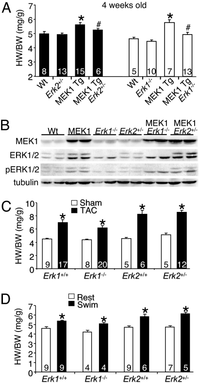
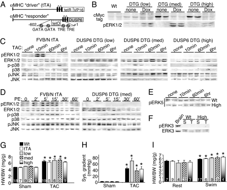
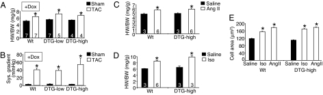
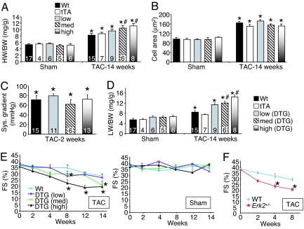
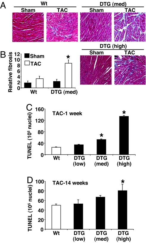
MAPK signaling pathways function as critical regulators of cellular differentiation, proliferation, stress responsiveness, and apoptosis. One branch of the MAPK signaling pathway that culminates in ERK1/2 activation is hypothesized to regulate the growth and adaptation of the heart to both physiologic and pathologic stimuli, given its known activation in response to virtually every stress- and agonist-induced hypertrophic stimulus examined to date. Here we investigated the requirement of ERK1/2 signaling in mediating the cardiac hypertrophic growth response in Erk1(-/-) and Erk2(+/-) mice, as well as in transgenic mice with inducible expression of an ERK1/2-inactivating phosphatase in the heart, dual-specificity phosphatase 6. Although inducible expression of dual-specificity phosphatase 6 in the heart eliminated ERK1/2 phosphorylation at baseline and after stimulation without affecting any other MAPK, it did not diminish the hypertrophic response to pressure overload stimulation, neuroendocrine agonist infusion, or exercise. Similarly, Erk1(-/-) and Erk2(+/-) mice showed no reduction in pathologic or physiologic stimulus-induced cardiac growth in vivo. However, blockade or deletion of cardiac ERK1/2 did predispose the heart to decompensation and failure after long-term pressure overload in conjunction with an increase in myocyte TUNEL. Thus, ERK1/2 signaling is not required for mediating physiologic or pathologic cardiac hypertrophy in vivo, although it does play a protective role in response to pathologic stimuli.


Conflict of interest statement

The authors declare no conflict of interest.

Figures

Fig. 1.
Fig. 1.
Erk1/2 gene-targeted mice undergo normal cardiac hypertrophy. (A) Heart weight/body weight (HW/BW) in the indicated groups of mice, assayed at 4 weeks of age. *, P < 0.05 vs. WT; #, P < 0.05 vs. MEK1 transgenic (Tg). (B) Western blot analysis of MEK1 protein, ERK1/2 protein, phospho-ERK1/2, and α-tubulin (control) from the hearts of the indicated mice (n = 2 blots). (C) Heart weight/body weight in the indicated groups of mice 2 weeks after TAC or a sham procedure. *, P < 0.05 vs. sham of each pairing. (D) Heart weight/body weight in the indicated groups of mice after 20 days of swimming exercise or rest. *, P < 0.05 vs. the rest of each pairing.
Fig. 2.
Fig. 2.
Characterization of cardiac-specific DUSP6-inducible transgenic mice. (A) Schematic of the bitransgenic inducible expression system used to regulate DUSP6 in the mouse heart. (B) Western blot for Myc-tagged DUSP6 protein in the hearts of the indicated mice on Dox (shutoff) or without Dox (induced). Corresponding basal ERK1/2 phosphorylation is also shown. (C) Western blot assessment of MAPK phosphorylation from hearts at baseline or after TAC for the indicated times in the different groups of adult mice without Dox (induced). At least six separate samples were analyzed in total per condition (although only two are shown). (D) Western blot assessment of cardiac MAPK phosphorylation at baseline or after acute systemic PE injection for the indicated times in mice without Dox (induced). The entire time course was repeated in separate mice with identical results. (E) Western blot of phosphorylated ERK5 (activated) in the hearts of WT or high-expressing DUSP6 DTG mice after 10 min, 60 min, or 6 h of TAC stimulation. (F) ERK3 phosphorylation was not detected in hearts of sham (S) or TAC (T) stimulated mice that were WT or high-expressing DUSP6 DTG, although ERK3 protein was detected. Brain extract was run as a control. (G) Heart weight/body weight in the indicated groups of mice 2 weeks after TAC or a sham procedure (no Dox, induced). *, P < 0.05 vs. sham of each pairing. (H) Systolic pressure gradient across the aortic constriction. *, P < 0.05 vs. sham. (I) Heart weight/body weight in the indicated groups of mice after 20 days of swimming or rest in the induced stated (no Dox). *, P < 0.05 vs. the rest of each pairing. The key in G applies to H and I as well.
Fig. 3.
Fig. 3.
Characterization of cardiac-specific DUSP6-inducible transgenic mice. (A) Heart weight/body weight in the indicated groups of mice on Dox (transgene off) 2 weeks after TAC or a sham procedure. *, P < 0.05 vs. sham of each pairing. (B) Corresponding systolic pressure gradients of the mice in A. (C) Heart weight/body weight in the indicated groups of mice 2 weeks after Ang II infusion or saline (no Dox, induced). *, P < 0.05 vs. saline. (D) Heart weight/body weight in the indicated groups of mice 2 weeks after Iso infusion or saline (no Dox, induced). *, P < 0.05 vs. saline. (E) Histological analysis of myocyte cross-sectional areas from ventricles of the indicated mice and indicated treatments (at least 500 myocytes were counted from three separate hearts in each group). *, P < 0.05 vs. saline.
Fig. 4.
Fig. 4.
Analysis of heart failure in DUSP6-inducible transgenic mice. (A) Heart weight/body weight in the indicated groups of mice 14 weeks after TAC or a sham procedure (no Dox, induced). *, P < 0.05 vs. sham of each pairing; #, P < 0.05 vs. WT or tTA TAC. (B) Histological analysis of myocyte cross-sectional areas from ventricles of the indicated mice and indicated treatments (at least 500 myocytes were counted from three separate hearts in each group). *, P < 0.05 vs. sham. (C) Corresponding systolic pressure gradients at 2 weeks of TAC across the aortic constriction for the mice shown in A. (D) Corresponding lung weight/body weight (LW/BW) for the mice in A. (E) Echocardiography assessment of fractional shortening (FS) in DUSP6 DTG mice after TAC (Left) or subjected to a sham procedure (Right) for the indicated times. *, P < 0.05 vs. WT. (F) Echocardiography assessment of fractional shortening (FS) in WT and Erk2+/− mice after TAC for the indicated time points. *, P < 0.05 vs. WT.
Fig. 5.
Fig. 5.
Histological assessment of fibrosis and TUNEL in DUSP6 DTG mice after TAC. (A) Representative Masson's trichrome-stained histological sections from the hearts of the indicated mice after 14 weeks of TAC or sham operation. (B) Quantitation of relative fibrosis of the hearts of medium-expressing DUSP6 DTG mice (n = 3 hearts each). (C) Measurement of TUNEL in cardiac histological sections from the indicated mice after 1 week of TAC stimulation (n = 3 hearts each, with at least 200,000 nuclei surveyed per heart). (D) Measurement of TUNEL in cardiac histological sections from the indicated mice after 14 weeks of TAC stimulation (n = 3 hearts each, with at least 200,000 nuclei surveyed per heart). *, P < 0.05 vs. WT TAC.

References

    1. Ho KK, Levy D, Kannel WB, Pinsky JL. J Am Coll Cardiol. 1993;22:6–13. - PubMed
    1. Levy D, Garrison RJ, Savage DD, Kannel WB, Castelli WP. N Engl J Med. 1990;322:1561–1566. - PubMed
    1. Dickhuth HH, Lehmann M, Auch-Schwelk W, Meinertz T, Keul J. J Cardiovasc Pharmacol. 1987;10:S71–S78. - PubMed
    1. Allen DL, Harrison BC, Maass A, Bell ML, Byrnes WC, Leinwand LA. J Appl Physiol. 2001;90:1900–1908. - PubMed
    1. Heineke J, Molkentin JD. Nat Rev Mol Cell Biol. 2006;7:589–600. - PubMed

Publication types

MeSH terms

Substances