. 2007 Aug 15;2(8):e754.
doi: 10.1371/journal.pone.0000754.

Biphasic Hoxd gene expression in shark paired fins reveals an ancient origin of the distal limb domain

Affiliations

Biphasic Hoxd gene expression in shark paired fins reveals an ancient origin of the distal limb domain

Renata Freitas et al. PLoS One. .

Abstract

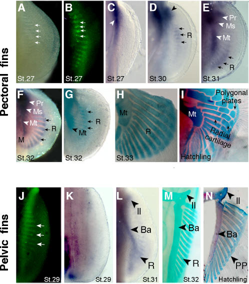
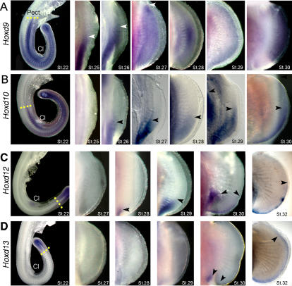
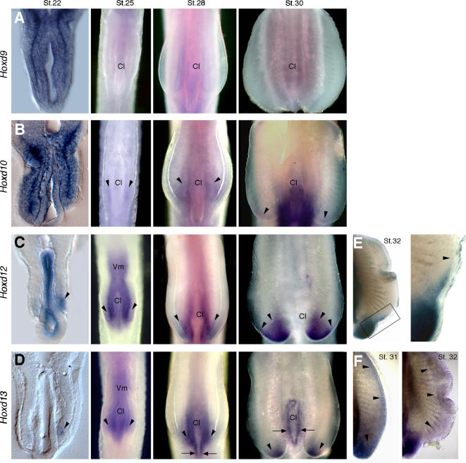
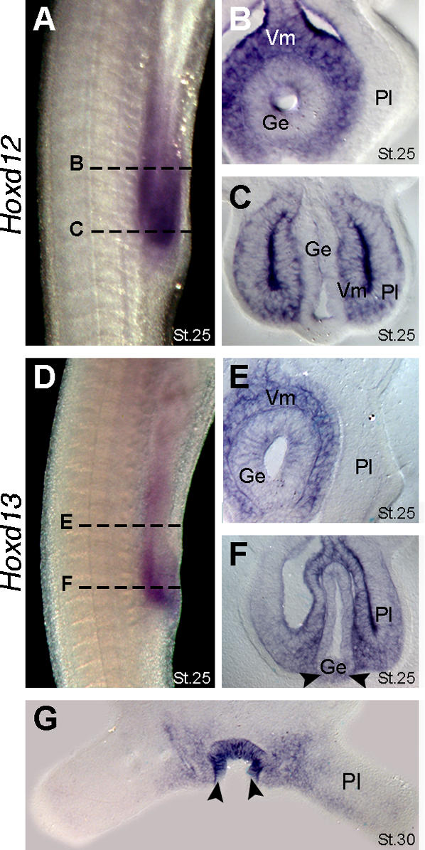
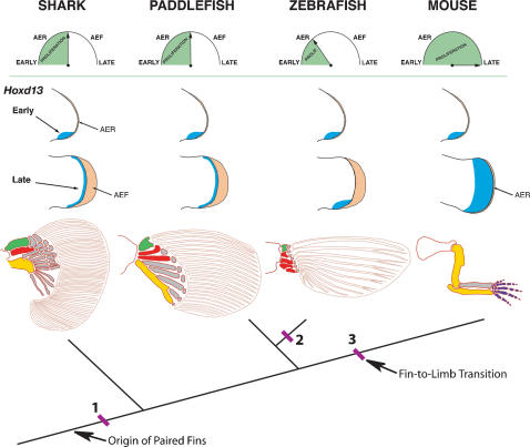
The evolutionary transition of fins to limbs involved development of a new suite of distal skeletal structures, the digits. During tetrapod limb development, genes at the 5' end of the HoxD cluster are expressed in two spatiotemporally distinct phases. In the first phase, Hoxd9-13 are activated sequentially and form nested domains along the anteroposterior axis of the limb. This initial phase patterns the limb from its proximal limit to the middle of the forearm. Later in development, a second wave of transcription results in 5' HoxD gene expression along the distal end of the limb bud, which regulates formation of digits. Studies of zebrafish fins showed that the second phase of Hox expression does not occur, leading to the idea that the origin of digits was driven by addition of the distal Hox expression domain in the earliest tetrapods. Here we test this hypothesis by investigating Hoxd gene expression during paired fin development in the shark Scyliorhinus canicula, a member of the most basal lineage of jawed vertebrates. We report that at early stages, 5'Hoxd genes are expressed in anteroposteriorly nested patterns, consistent with the initial wave of Hoxd transcription in teleost and tetrapod paired appendages. Unexpectedly, a second phase of expression occurs at later stages of shark fin development, in which Hoxd12 and Hoxd13 are re-expressed along the distal margin of the fin buds. This second phase is similar to that observed in tetrapod limbs. The results indicate that a second, distal phase of Hoxd gene expression is not uniquely associated with tetrapod digit development, but is more likely a plesiomorphic condition present the common ancestor of chondrichthyans and osteichthyans. We propose that a temporal extension, rather than de novo activation, of Hoxd expression in the distal part of the fin may have led to the evolution of digits.


Conflict of interest statement

Competing Interests: The authors have declared that no competing interests exist.

Figures

Figure 1
Figure 1. Endoskeletal development in catshark pectoral and pelvic fins.
Ventral views of pectoral (A–I) and pelvic (J–N) fins. Stages (St.) of development indicated at bottom of each panel. (A) Light micrograph of pectoral fin showing gaps in the pectoral fin plate. (B) Acridine orange staining (green fluorescence) shows apoptotic cells in the gaps observed in panel A. Arrows in A and B mark four examples. (C) Sox8 expression marks initiation of chondrogenesis in the pectoral girdle region (arrowhead). Note absence of chondrogenesis in the fin plate at this stage. (D) Sox8 expression marks initiation of chondrogenesis in anterior part of the fin plate, in basal cartilages (arrowhead) and radials (arrows). (E) Sox8 domain prefigures development of the basal cartilages along the anteroposterior axis of the fin: Pr, propterygium; Ms, mesopterygium; Mt, metapterygium; R, radials. Arrows mark expression in the most posterior radials. (F) Sox8 expression in basal cartilages (arrowheads) and in all radials along the anteroposterior axis (subset of radials marked with arrows). (G, H) Alcian green staining of pectoral fins. Note that radials chondrify in domains pre-established by Sox8 expression domains (compare with panels F and G). Chondrified, unsegmented radials are seen in H. (I) Alcian blue and alizarin red stained pectoral fin showing a fully developed cartilaginous endoskeleton at the time of hatching. Note segmentation of proximal radials, intermediate radials and distal polygonal plates (compare panels H and I). (J) Acridine orange-positive cells in gaps of the pelvic fin plate. (K) Sox8 expression marks initiation of chondrogenesis proximal, posterior region of fin. Note absence of chondrogenesis in the fin plate at this stage. (L) Sox8 expression prefigures development of endoskeletal elements in the pelvic fin. Il, iliac process; Ba, basipterygium; R, radials. (M) Alcian green staining of the pelvic fin showing chondrified unsegmented radials. (N) Alcian blue and alizarin red staining of the pelvic fin showing fully developed cartilaginous endoskeleton at hatching. Note segmentation of the radials into distal polygonal plates (PP) and proximal radials (compare panels M and N).
Figure 2
Figure 2. Expression of Hoxd genes in catshark pectoral fins.
Stages of development indicated in lower right corners of each panel. (A–D) Whole mount in situ hybridizations showing expression of Hoxd9 (A), Hoxd10 (B), Hoxd12 (C) and Hoxd13 (D). Pect, Pectoral fin bud; Cl, cloaca. Note anterior expansion of Hoxd12 and Hoxd13 in distal fin at stage 32. Arrows mark anterior limits of expression. Yellow dotted lines in the left column mark the anterior boundaries of expression at stage 22.
Figure 3
Figure 3. Expression of Hoxd genes in catshark pelvic fins.
Stages of development indicated in the top of each column in A–D and in upper right corner in E and F. Left column shows transverse histological sections at level of cloaca (Cl) and pelvic fins. All other panels show whole mounts in ventral view. (A–D) Whole mount in situ hybridizations showing expression of Hoxd9 (A), Hoxd10 (B), Hoxd12 (C) and Hoxd13 (D). Arrowheads mark expression in pelvic fin buds. Arrows in D mark expression in cloacal epithelium. (E, F) Pelvic fins showing expression of Hoxd12 at stage 32 (E) and Hoxd13 at stages 31 and 32 (F). Boxed area in E is shown in high magnification at right. Arrowheads in E mark anterior limits of expression, and in F they outline the extent of the distal Hoxd13 domain.
Figure 4
Figure 4. Expression of Hoxd12 and Hoxd13 in the cloacal region of catsharks.
(A) Lateral view of pelvic fin region showing Hoxd12 expression at stage 25. Dashed lines mark the approximate planes of section showed in panels B and C. (B) Transverse section showing Hoxd12 expression in visceral mesoderm (Vm) and gut endoderm (Ge). Note absence of Hoxd12 expression in anterior part of the pelvic fin (Pl). (C) Transverse section showing Hoxd12 expression in the posterior part of pelvic fin and adjacent visceral mesoderm. Note absence of Hoxd12 expression in the gut endoderm. (D) Lateral view of the pelvic fin region showing Hoxd13 expression at stage 25. Note that Hoxd13 domain lies posterior to Hoxd12 domain (compare with panel A). Dashed lines mark the approximate planes of the section showed in panels E and F. (E) Transverse section showing Hoxd13 expression in the visceral mesoderm and gut endoderm. Note absence of Hoxd13 expression in the anterior part of pelvic fin. (F) Transverse section showing Hoxd13 expression in the posterior part of the fin, visceral mesoderm and ventral endoderm. Arrowheads mark expression in endoderm (contrast with absence of Hoxd12 in endoderm in panel C). (G) Transverse section throughout the pelvic fins at stage 30 showing Hoxd13 expression in the cloacal epithelium (arrowheads).
Figure 5
Figure 5. Model for the origin of digits by temporal extension of distal Hoxd13 expression.
Tree shows phylogenetic relationships of shark, paddlefish, zebrafish and mouse. Top row shows hypothetical timing for the transition of the apical ectodermal ridge (AER) to apical ectodermal fold (AEF); note that the mouse maintains an AER and does not form an AEF. Green shading represents proliferative period for endoskeletal progenitor cells. Middle rows show Hoxd13 expression domains (blue) at early and late stages of fin and limb bud outgrowth. AER and AEF are shaded orange. Bottom row shows pectoral appendicular skeleton for each taxon. Endoskeletal bones are shaded as follows: green, propterygium; red, mesopterygium, yellow, metapterygium. Dermal fin rays are shown as unshaded elements within fin blade. The model suggests that a second phase of distal Hoxd13 expression was present in the paired fins of the common ancestor of chondrichthyans and osteichthyans (at position 1), and that loss of the distal Hoxd13 domain in teleosts (position 2) and its spatial expansion in tetrapods (position 3) may have been associated with temporal modulation of endoskeletal progenitor cell proliferation. Early conversion of the AER to an AEF would be expected to truncate or eliminate phase II expression of Hoxd13 and reduce the fin endoskeleton, as seen in zebrafish, whereas prolonged signaling by the AER would be expected to extend Phase II and expand the Hoxd13 domain, giving rise to digits in the tetrapod lineage. Clock model after ; skeletal patterns after , , , .

References

    1. Coates MI. The evolution of paired fins. Theory in Biosciences. 2003;122:266–287.
    1. Shubin NH, Daeschler EB, Jenkins FA., Jr The pectoral fin of Tiktaalik roseae and the origin of the tetrapod limb. Nature. 2006;440:764–771. - PubMed
    1. Zakany J, Fromental-Ramain C, Warot X, Duboule D. Regulation of number and size of digits by posterior Hox genes: a dose-dependent mechanism with potential evolutionary implications. Proc Natl Acad Sci U S A. 1997;94:13695–13700. - PMC - PubMed
    1. Grandel H. Approaches to a comparison of fin and limb structure and development. Theory Biosci. 2003;122:288–301.
    1. Zhang XG, Hou XG. Evidence for a single median fin-fold and tail in the Lower Cambrian vertebrate, Haikouichthys ercaicunensis. J Evol Biol. 2004;17:1162–1166. - PubMed

Publication types

Substances