

Oncogene MYCN regulates localization of NKT cells to the site of disease in neuroblastoma
- PMID: 17710228
- PMCID: PMC1940236
- DOI: 10.1172/JCI30751
Oncogene MYCN regulates localization of NKT cells to the site of disease in neuroblastoma
Abstract
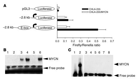
Valpha24-invariant natural killer T (NKT) cells are potentially important for antitumor immunity. We and others have previously demonstrated positive associations between NKT cell presence in primary tumors and long-term survival in distinct human cancers. However, the mechanism by which aggressive tumors avoid infiltration with NKT and other T cells remains poorly understood. Here, we report that the v-myc myelocytomatosis viral related oncogene, neuroblastoma derived (MYCN), the hallmark of aggressive neuroblastoma, repressed expression of monocyte chemoattractant protein-1/CC chemokine ligand 2 (MCP-1/CCL2), a chemokine required for NKT cell chemoattraction. MYCN knockdown in MYCN-amplified neuroblastoma cell lines restored CCL2 production and NKT cell chemoattraction. Unlike other oncogenes, MYCN repressed chemokine expression in a STAT3-independent manner, requiring an E-box element in the CCL2 promoter to mediate transcriptional repression. MYCN overexpression in neuroblastoma xenografts in NOD/SCID mice severely inhibited their ability to attract human NKT cells, T cells, and monocytes. Patients with MYCN-amplified neuroblastoma metastatic to bone marrow had 4-fold fewer NKT cells in their bone marrow than did their nonamplified counterparts, indicating that the MYCN-mediated immune escape mechanism, which we believe to be novel, is operative in metastatic cancer and should be considered in tumor immunobiology and for the development of new therapeutic strategies.
Figures


Similar articles
-
MYCN enhances P-gp/MDR1 gene expression in the human metastatic neuroblastoma IGR-N-91 model.Am J Pathol. 2003 Jul;163(1):321-31. doi: 10.1016/S0002-9440(10)63656-5. Am J Pathol. 2003. PMID: 12819037 Free PMC article.
-
Cellular retinoic acid-binding protein II is a direct transcriptional target of MycN in neuroblastoma.Cancer Res. 2006 Aug 15;66(16):8100-8. doi: 10.1158/0008-5472.CAN-05-4519. Cancer Res. 2006. PMID: 16912187
-
GRHL1 acts as tumor suppressor in neuroblastoma and is negatively regulated by MYCN and HDAC3.Cancer Res. 2014 May 1;74(9):2604-16. doi: 10.1158/0008-5472.CAN-13-1904. Epub 2014 Jan 13. Cancer Res. 2014. PMID: 24419085
-
MDM2 as MYCN transcriptional target: implications for neuroblastoma pathogenesis.Cancer Lett. 2005 Oct 18;228(1-2):21-7. doi: 10.1016/j.canlet.2005.01.050. Cancer Lett. 2005. PMID: 15927364 Review.
-
The MYCN oncoprotein as a drug development target.Cancer Lett. 2003 Jul 18;197(1-2):125-30. doi: 10.1016/s0304-3835(03)00096-x. Cancer Lett. 2003. PMID: 12880971 Review.
Cited by
-
Altered Lipid Tumor Environment and Its Potential Effects on NKT Cell Function in Tumor Immunity.Front Immunol. 2019 Sep 18;10:2187. doi: 10.3389/fimmu.2019.02187. eCollection 2019. Front Immunol. 2019. PMID: 31620124 Free PMC article. Review.
-
Adoptive cell therapy in paediatric extracranial solid tumours: current approaches and future challenges.Eur J Cancer. 2023 Nov;194:113347. doi: 10.1016/j.ejca.2023.113347. Epub 2023 Sep 18. Eur J Cancer. 2023. PMID: 37832507 Free PMC article. Review.
-
Immunology and immunotherapy of neuroblastoma.Semin Cancer Biol. 2011 Oct;21(4):229-37. doi: 10.1016/j.semcancer.2011.09.012. Epub 2011 Sep 28. Semin Cancer Biol. 2011. PMID: 21971567 Free PMC article. Review.
-
Invariant NKT cells with chimeric antigen receptor provide a novel platform for safe and effective cancer immunotherapy.Blood. 2014 Oct 30;124(18):2824-33. doi: 10.1182/blood-2013-11-541235. Epub 2014 Jul 21. Blood. 2014. PMID: 25049283 Free PMC article.
-
MYC regulates metabolism through vesicular transfer of glycolytic kinases.Open Biol. 2021 Dec;11(12):210276. doi: 10.1098/rsob.210276. Epub 2021 Dec 1. Open Biol. 2021. PMID: 34847775 Free PMC article.
References
-
- Kronenberg M. Toward an understanding of NKT cell biology: progress and paradoxes. Annu. Rev. Immunol. 2005;23:877–900. - PubMed
-
- Van Kaer L., Joyce S. Innate immunity: NKT cells in the spotlight. Curr. Biol. 2005;15:R429–R431. - PubMed
-
- Swann J., Crowe N.Y., Hayakawa Y., Godfrey D.I., Smyth M.J. Regulation of antitumour immunity by CD1d-restricted NKT cells. Immunol. Cell Biol. 2004;82:323–331. - PubMed
-
- Gumperz J.E. CD1d-restricted “NKT” cells and myeloid IL-12 production: an immunological crossroads leading to promotion or suppression of effective anti-tumor immune responses? J. Leukoc. Biol. 2004;76:307–313. - PubMed
-
- Yanagisawa K., et al. Impaired proliferative response of V alpha 24 NKT cells from cancer patients against alpha-galactosylceramide. J. Immunol. 2002;168:6494–6499. - PubMed
Publication types
MeSH terms
Substances
Grants and funding
LinkOut - more resources
Full Text Sources
Other Literature Sources
Medical
Molecular Biology Databases
Research Materials
Miscellaneous
