Xanthogranulomatous endometritis: a challenging imitator of endometrial carcinoma
- PMID: 17710239
- PMCID: PMC1939916
- DOI: 10.1155/2007/34763
Xanthogranulomatous endometritis: a challenging imitator of endometrial carcinoma
Abstract
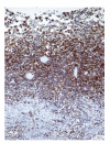
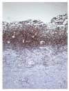
Xanthogranulomatous inflammation is a distinguished histopathological entity affecting several organs, predominantly the kidney and gallbladder. So far, only a small number of cases of xanthogranulomatous inflammation occurring in female genital tract have been described, most frequently affecting the endometrium and histologically characterized by replacement of endometrium by xanthogranulomatous inflammation composed of abundant foamy histiocytes, siderophages, giant cells, fibrosis, calcification and accompanying polymorphonuclear leucocytes, plasma cells and lymphocytes of polyclonal origin. We present a case of a 69-year-old female complained of post menopausal bleeding and weight loss. Clinical preliminary diagnoses were endometrial carcinoma or hyperplasia and ultrasound was supposed to be endometrial malignancy, hyperplasia or pyometra by radiologist. Histopathological examination of uterus revealed xanthogranulomatous endometritis. Since xanthogranulomatous endometritis may mimic endometrial malignancy clinically and pathologically as a result of the replacement of the endometrium and occasionally invasion of the myometrium by friable yellowish tissue composed of histiocytes, knowledge of this unusual inflammatory disease is needed for both clinicians and pathologists.
Figures


References
-
- Pounder DJ, Iyer PV. Xanthogranulomatous endometritis associated with endometrial carcinoma. Archives of Pathology and Laboratory Medicine. 1985;109(1):73–75. - PubMed
-
- Russack V, Lammers RJ. Xanthogranulomatous endometritis. Report of six cases and a proposed mechanism of development. Archives of Pathology and Laboratory Medicine. 1990;114(9):929–932. - PubMed
-
- Ashkenazy M, Lancet M, Borenstein R, Czernobilsky B. Endometrial foam cells. Non-estrogenic and estrogenic. Acta Obstetricia et Gynecologica Scandinavica. 1983;62(3):193–197. - PubMed
-
- Barua R, Krikland JA, Petrucco OM. Xanthogranulomatous endometritis: case report. Pathology. 1978;10(2):161–164. - PubMed
-
- Noack F, Briese J, Stellmacher F, Hornung D, Horny H-P. Lethal outcome in xanthogranulomatous endometritis. APMIS. 2006;114(5):386–388. - PubMed
Publication types
MeSH terms
LinkOut - more resources
Full Text Sources
Medical

