. 2007 Oct;51(4):499-508.
doi: 10.1111/j.1365-2559.2007.02791.x. Epub 2007 Aug 17.

Increased cytoplasmic level of migfilin is associated with higher grades of human leiomyosarcoma

Affiliations

Increased cytoplasmic level of migfilin is associated with higher grades of human leiomyosarcoma

D J Papachristou et al. Histopathology. 2007 Oct.

Abstract

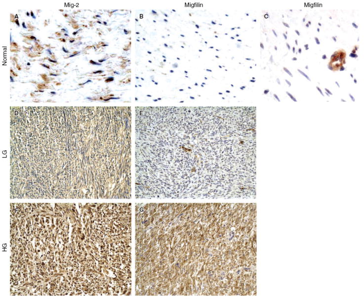
Aims: Leiomyosarcomas (LMS) are malignant neoplasms composed of cells that exhibit distinct smooth muscle differentiation. The molecular and cytogenetic features of LMS are complex and no consistent aberrations have been reported to date. Mitogen inducible gene-2 (Mig-2), kindlin and migfilin are recently identified cell-matrix adhesion proteins. The aim was to determine the expression and distribution of these proteins in human smooth muscle tumours of somatic soft tissue.

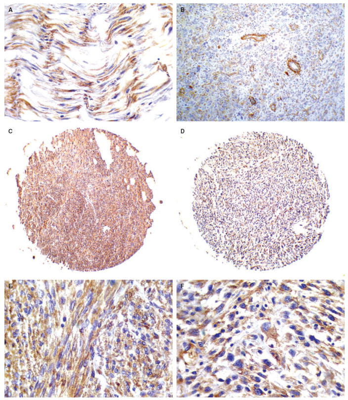
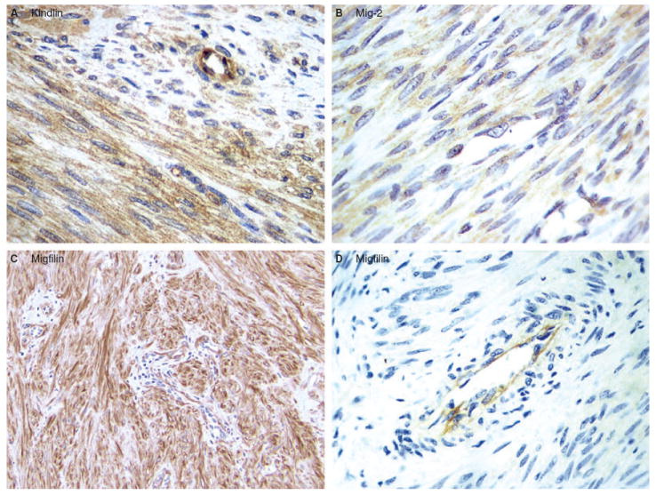
Methods and results: Immunohistochemistry was performed on a human LMS tissue microarray and on sections of human leiomyomas (LM) and normal smooth muscle. Migfilin was barely detectable in normal smooth muscle cells, whereas increased levels of migfilin were observed in the majority of LM and LMS. Furthermore, the cytoplasmic level of migfilin was strongly associated with higher tumour grades. Additionally, the cytoplasmic levels of migfilin and Mig-2 were correlated with each other, suggesting an association between the two in the cytoplasm. Kindlin was expressed in normal smooth muscle, LM and LMS, and its level did not correlate with tumour grade.

Conclusions: Our results suggest a role for cytoplasmic migfilin in the progression of LMS and identify cytoplasmic migfilin as a potentially important biological marker for human LMS progression.


Figures

Figure 1
Figure 1
RNAi suppression of kindlin expression in human keratinocytes. Human HaCaT keratinocytes transfected with the kindlin small interfering (si) RNA (lane 1) or a control RNA (lane 2) were probed with monoclonal anti-kindlin antibody 4A5 (top panel) or an anti-actin antibody (bottom panel) as indicated. Note that the amount of kindlin-1 was substantially reduced in the kindlin siRNA transfectants (compare lanes 1 and 2, top panel).
Figure 2
Figure 2
Localization of kindlin at cell–matrix adhesion sites in human keratinocytes. HaCat cells were stained with either the anti-kindlin monoclonal antibody (clone 4A5.14) and a Rhodamine Red™-conjugated goat antimouse IgG secondary antibody (B), or just the secondary antibody (A), which served as a negative control. (Leica DM R fluorescence microscope. Bar in panel B = 8 μm.)
Figure 3
Figure 3
Expression of kindlin in normal smooth muscle, low- and high-grade leiomyosarcoma (LMS). A, Normal smooth muscle cells display immunopositivity for kindlin. B, Moderate immunopositivity for kindlin (2+) in a high-grade LMS. Endothelial cells of tumour vessels are strongly positive for kindlin. C,E, Strong kindlin immunopositivity (3+) in a low-grade LMS. D,F, High-grade LMS exhibiting strong immunoreactivity (3+) for kindlin.
Figure 4
Figure 4
Expression of kindlin, Mig-2 and migfilin in leiomyoma (LM). A, Kindlin expression. Kindlin was detected in the majority (89%) of the LM cases. A representative diffuse, cytoplasmic kindlin staining of LM is shown. B, Mig-2 expression. Diffuse, cytoplasmic Mig-2 was detected in all of the LM cases. Representative Mig-2 reactivity is shown. C,D, Migfilin expression. Migfilin was detected in 14 out of 21 (66.7%) LM cases. C, Strongly positive, primarily cytoplasmic migfilin reactivity in a case of soft tissue LM. D, Negative migfilin reactivity in a case of soft tissue LM. Note that strong migfilin immunoreactivity was detected in endothelial cells (D).
Figure 5
Figure 5
Expression of Mig-2 and migfilin in normal smooth muscle, low- and high-grade leiomyosarcoma (LMS). A, Normal smooth muscle cells exhibit positive nuclear and cytoplasmic immunoreactivity for Mig-2. B, Normal smooth muscle cells exhibit no immunoreactivity for migfilin. C, In normal soft tissue sections containing blood vessels, a high level of migfilin was detected in endothelial cells. D, Mig-2 exhibits moderate cytoplasmic immunoreactivity (2+) in a low-grade LMS. E, Low-grade LMS exhibiting weak immunopositivity for migfilin (1+). Endothelial cells show strong expression of migfilin. F, Strong nuclear and cytoplasmic immunoreactivity of Mig-2 (3+) in a high-grade LMS. G, High-grade LMS showing strong (3+), nuclear and cytoplasmic immunopositivity for migfilin. See Table 1 for a summary of tumour grade, clinical features and immunopositivity/localization of migfilin and Mig-2 in all 40 human LMS that were analysed.

References

    1. Enzinger F, Weis SW. Leiomyosarcoma. In: Weis SW, Goldblum JR, editors. Soft tissue tumors. 4. Mosby; St Louis: 2001. pp. 724–748.
    1. Evans H, Shipley J. Leiomyosarcoma. In: Fletcher CDM, Unni KK, Mertens F, editors. World Health Organization classification of tumors. Pathology and genetics of tumors of soft tissue and bone. Lyon: IARC Press; 2002. pp. 131–134.
    1. Ren B, Yu YP, Jing L, et al. Gene expression analysis of human soft tissue leiomyosarcomas. Hum Pathol. 2003;34:549–558. - PubMed
    1. Sepulveda J, Gkretsi V, Wu C. Assembly and signaling of the cell–extracellular matrix adhesion complexes. Curr Top Dev Biol. 2005;68:183–225. - PubMed
    1. Jamora C, Fuchs E. Intercellular adhesion, signalling and the cytoskeleton. Nat Cell Biol. 2002;4:E101–108. - PubMed

Publication types

MeSH terms