

Epidermal stem cells are defined by global histone modifications that are altered by Myc-induced differentiation
- PMID: 17712411
- PMCID: PMC1945016
- DOI: 10.1371/journal.pone.0000763
Epidermal stem cells are defined by global histone modifications that are altered by Myc-induced differentiation
Abstract
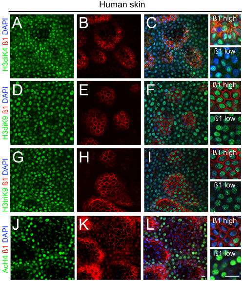
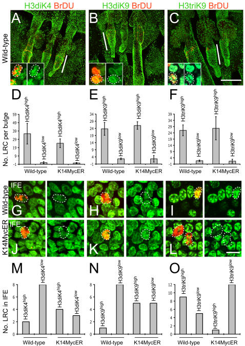
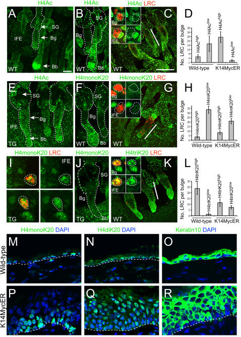
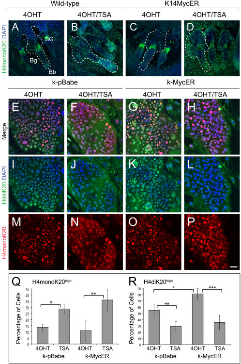
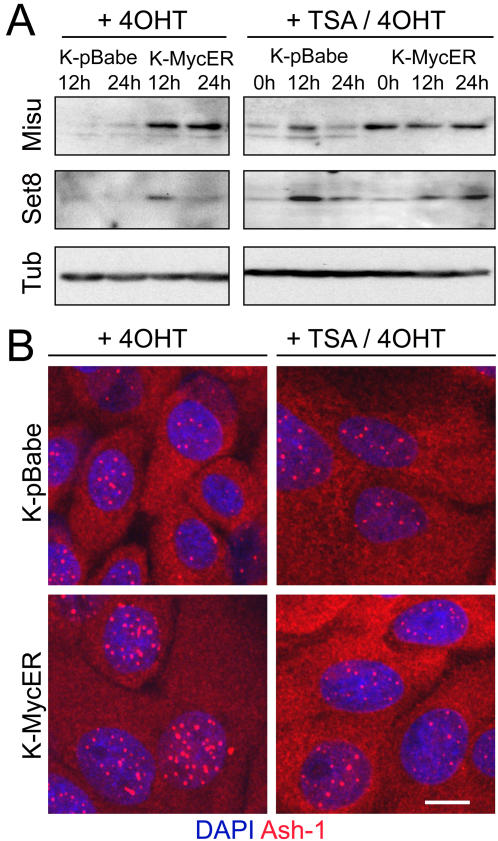
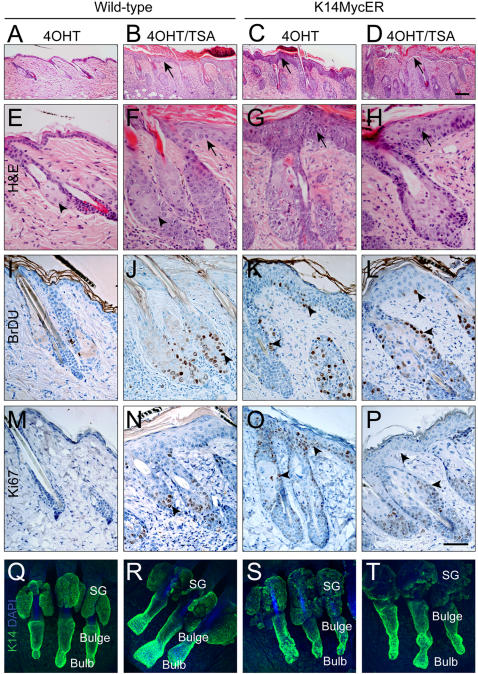
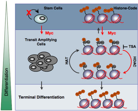
Activation of Myc induces epidermal stem cells to exit their niche and differentiate into sebocytes and interfollicular epidermis, a process that is associated with widespread changes in gene transcription. We have identified chromatin modifications that are characteristic of epidermal stem cells and investigated the effects of Myc activation. Quiescent stem cells in the interfollicular epidermis and the hair follicle bulge had high levels of tri-methylated histone H3 at lysine 9 and H4 at lysine 20. Chromatin in both stem cell populations was hypoacteylated at histone H4 and lacked mono-methylation of histone H4 at lysine 20. Myc-induced exit from the stem cell niche correlated with increased acetylation at histone H4 and transiently increased mono-methylation at lysine 20. The latter was replaced by epigenetic modifications that are largely associated with chromatin silencing: di-methylation at histone H3 lysine 9 and histone H4 lysine 20. These modifications correlated with changes in the specific histone methyltransferases Set8 and Ash-1. The Myc-induced switch from mono- to di-methylated H4K20 required HDAC activity and was blocked by the HDAC inhibitor trichostatin A (TSA). TSA treatment induced a similar epidermal phenotype to activation of Myc, and activation of Myc in the presence of TSA resulted in massive stimulation of terminal differentiation. We conclude that Myc-induced chromatin modifications play a major role in Myc-induced exit from the stem cell compartment.
Conflict of interest statement
Figures
Similar articles
-
c-Myc activation in transgenic mouse epidermis results in mobilization of stem cells and differentiation of their progeny.Curr Biol. 2001 Apr 17;11(8):558-68. doi: 10.1016/s0960-9822(01)00154-3. Curr Biol. 2001. PMID: 11369200
-
Inhibition of histone deacetylases by Trichostatin A leads to a HoxB4-independent increase of hematopoietic progenitor/stem cell frequencies as a result of selective survival.Cytotherapy. 2010 Nov;12(7):899-908. doi: 10.3109/14653240903580254. Cytotherapy. 2010. PMID: 20210674
-
Coordinated changes in DNA methylation and histone modifications regulate silencing/derepression of luteinizing hormone receptor gene transcription.Mol Cell Biol. 2005 Sep;25(18):7929-39. doi: 10.1128/MCB.25.18.7929-7939.2005. Mol Cell Biol. 2005. PMID: 16135786 Free PMC article.
-
Epigenetic landscape of amphetamine and methamphetamine addiction in rodents.Epigenetics. 2015;10(7):574-80. doi: 10.1080/15592294.2015.1055441. Epigenetics. 2015. PMID: 26023847 Free PMC article. Review.
-
MYC in mammalian epidermis: how can an oncogene stimulate differentiation?Nat Rev Cancer. 2008 Mar;8(3):234-42. doi: 10.1038/nrc2328. Nat Rev Cancer. 2008. PMID: 18292777 Free PMC article. Review.
Cited by
-
Cells, growth factors and biomaterials used in tissue engineering for hair follicles regeneration.Regen Ther. 2022 Nov 25;21:596-610. doi: 10.1016/j.reth.2022.11.003. eCollection 2022 Dec. Regen Ther. 2022. PMID: 36475027 Free PMC article. Review.
-
Epigenetic responses to environmental change and their evolutionary implications.Philos Trans R Soc Lond B Biol Sci. 2009 Nov 27;364(1534):3403-18. doi: 10.1098/rstb.2009.0125. Philos Trans R Soc Lond B Biol Sci. 2009. PMID: 19833651 Free PMC article. Review.
-
Dose and context dependent effects of Myc on epidermal stem cell proliferation and differentiation.EMBO Mol Med. 2010 Jan;2(1):16-25. doi: 10.1002/emmm.200900047. EMBO Mol Med. 2010. PMID: 20043278 Free PMC article.
-
Epigenetic and metabolic regulation of epidermal homeostasis.Exp Dermatol. 2021 Aug;30(8):1009-1022. doi: 10.1111/exd.14305. Epub 2021 Mar 10. Exp Dermatol. 2021. PMID: 33600038 Free PMC article. Review.
-
Role of histone deacetylase 2 in epigenetics and cellular senescence: implications in lung inflammaging and COPD.Am J Physiol Lung Cell Mol Physiol. 2012 Oct 1;303(7):L557-66. doi: 10.1152/ajplung.00175.2012. Epub 2012 Jul 27. Am J Physiol Lung Cell Mol Physiol. 2012. PMID: 22842217 Free PMC article. Review.
References
-
- Jenuwein T, Allis CD. Translating the histone code. Science. 2001;293:1074–1080. - PubMed
-
- Lund AH, van Lohuizen M. Epigenetics and cancer. Genes Dev. 2004;18:2315–2335. - PubMed
-
- Hsieh J, Gage FH. Chromatin remodeling in neural development and plasticity. Curr Opin Cell Biol. 2005;17:664–671. - PubMed
-
- Xi R, Xie T. Stem cell self-renewal controlled by chromatin remodeling factors. Science. 2005;310:1487–1489. - PubMed
-
- Kondo T. Epigenetic alchemy for cell fate conversion. Curr Opin Genet Dev 2006 - PubMed
Publication types
MeSH terms
Substances
Grants and funding
LinkOut - more resources
Full Text Sources
Medical
Research Materials
Miscellaneous
