

Low numbers of FOXP3 positive regulatory T cells are present in all developmental stages of human atherosclerotic lesions
- PMID: 17712427
- PMCID: PMC1945014
- DOI: 10.1371/journal.pone.0000779
Low numbers of FOXP3 positive regulatory T cells are present in all developmental stages of human atherosclerotic lesions
Abstract
Background: T cell mediated inflammation contributes to atherogenesis and the onset of acute cardiovascular disease. Effector T cell functions are under a tight control of a specialized T cell subset, regulatory T cells (Treg). At present, nothing is known about the in situ presence of Treg in human atherosclerotic tissue. In the present study we investigated the frequency of naturally occurring Treg cells in all developmental stages of human atherosclerotic lesions including complicated thrombosed plaques.
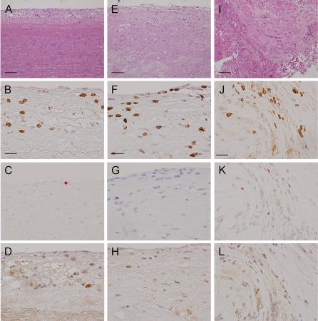
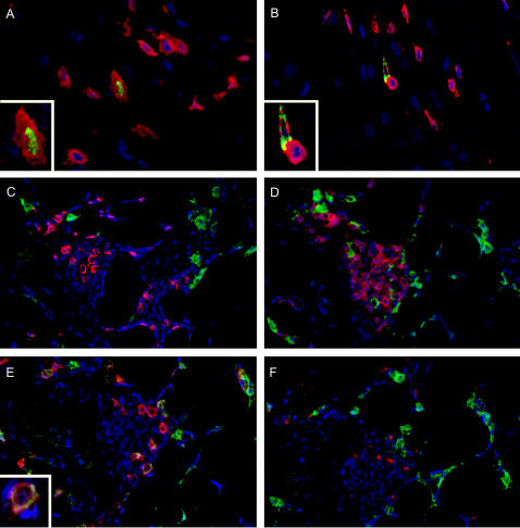
Methodology: Normal arteries, early lesions (American Heart Association classification types I, II, and III), fibrosclerotic plaques (types Vb and Vc) and 'high risk' plaques (types IV, Va and VI) were obtained at surgery and autopsy. Serial sections were immunostained for markers specific for regulatory T cells (FOXP3 and GITR) and the frequency of these cells was expressed as a percentage of the total numbers of CD3+ T cells. Results were compared with Treg counts in biopsies of normal and inflammatory skin lesions (psoriasis, spongiotic dermatitis and lichen planus).
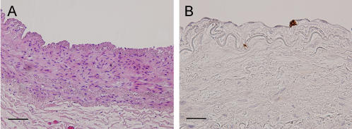
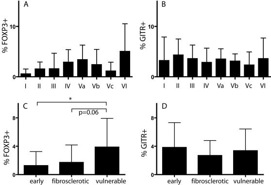
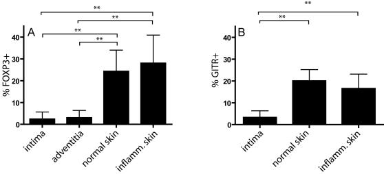
Principle findings: In normal vessel fragments T cells were virtually absent. Treg were present in the intima during all stages of plaque development (0.5-5%). Also in the adventitia of atherosclerotic vessels Treg were encountered, in similar low amounts. High risk lesions contained significantly increased numbers of Treg compared to early lesions (mean: 3.9 and 1.2%, respectively). The frequency of FOXP3+ cells in high risk lesions was also higher compared to stable lesions (1.7%), but this difference was not significant. The mean numbers of intimal FOXP3 positive cells in atherosclerotic lesions (2.4%) was much lower than those in normal (24.3%) or inflammatory skin lesions (28%).
Conclusion: Low frequencies of Treg in all developmental stages of human plaque formation could explain the smoldering chronic inflammatory process that takes place throughout the longstanding course of atherosclerosis.
Conflict of interest statement
Figures
References
-
- Ross R. Atherosclerosis–an inflammatory disease. N Engl J Med. 1999;340:115–126. - PubMed
-
- Jonasson L, Holm J, Skalli O, Bondjers G, Hansson GK. Regional accumulations of T cells, macrophages, and smooth muscle cells in the human atherosclerotic plaque. Arteriosclerosis. 1986;6:131–138. - PubMed
-
- van der Wal AC, Das PK, Bentz van de Berg D, van der Loos CM, Becker AE. Atherosclerotic lesions in humans. In situ immunophenotypic analysis suggesting an immune mediated response. Laboratory Investigation. 1989;61:166–170. - PubMed
-
- Kovanen PT, Kaartinen M, Paavonen T. Infiltrates of activated mast cells at the site of coronary atheromatous erosion or rupture in myocardial infarction. Circulation. 1995;92:1084–1088. - PubMed
-
- Hosono M, de Boer OJ, van der Wal AC, van der Loos CM, Teeling P, et al. Increased expression of T cell activation markers (CD25, CD26, CD40L and CD69) in atherectomy specimens of patients with unstable angina and acute myocardial infarction. Atherosclerosis. 2003;168:73–80. - PubMed
Publication types
MeSH terms
Substances
LinkOut - more resources
Full Text Sources
Other Literature Sources
Medical
