

Review
doi: 10.1155/2007/52487.
Pharmacotherapies for diabetic retinopathy: present and future
Affiliations
- PMID: 17713597
- PMCID: PMC1940315
- DOI: 10.1155/2007/52487
Item in Clipboard
Review
Pharmacotherapies for diabetic retinopathy: present and future
Exp Diabetes Res.
2007.
Abstract
Diabetic retinopathy remains a major cause of worldwide preventable blindness. Measures to avoid blindness include medical management (control of blood sugar, blood pressure, and serum lipids) and ocular management (laser photocoagulation and pars plana vitrectomy). Adjunctive pharmacologic therapies (intravitreal triamcinolone acetonide and anti-vascular endothelial growth factor agents) have shown early promise in the treatment of both diabetic macular edema and proliferative diabetic retinopathy. Other medications under investigation include the fluocinolone acetonide implantable device, extended-release dexamethasone implant, oral ruboxistaurin, and intravitreal hyaluronidase.
Figures
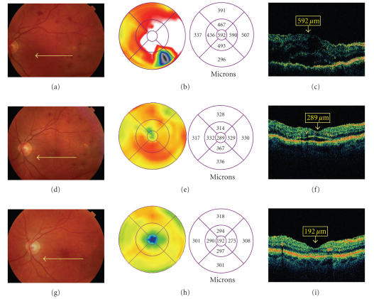
Intravitreal triamcinolone acetonide for diabetic macular edema. A patient presented with diabetic macular edema, visual acuity 20/60. Fundus photography (a) and optical coherence tomography (OCT) (b, c) are shown. The patient was treated with intravitreal triamcinolone acetonide. One month after treatment, visual acuity improved to 20/40, with improvement of macular edema on photography (d) and OCT (e, f). Four months after treatment, visual acuity improved to 20/20, with further improvement of macular edema on photography (g)
and OCT (h, i).
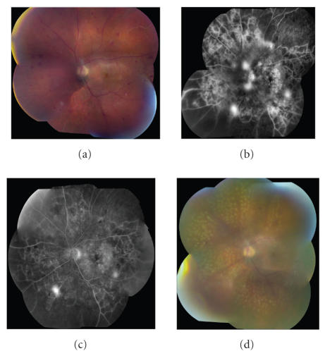
Intravitreal bevacizumab for proliferative diabetic retinopathy. A patient presented with proliferative diabetic retinopathy. Fundus photography (a) and fluorescein angiography (b) are shown. The patient was treated with intravitreal bevacizumab. Followup fluorescein angiography demonstrated improvement in angiographic leakage (c). Panretinal photocoagulation was then applied (d). (Case courtesy of Geeta Lalwani, MD, and Carmen A. Puliafito, MD, MBA.)
Similar articles
-
Update on treatments for diabetic macular edema.Curr Opin Ophthalmol. 2008 May;19(3):185-9. doi: 10.1097/ICU.0b013e3282fb7c45. Curr Opin Ophthalmol. 2008. PMID: 18408491 Review.
-
Changing paradigms in the treatment of diabetic retinopathy.Curr Opin Ophthalmol. 2009 Nov;20(6):532-8. doi: 10.1097/ICU.0b013e328330b533. Curr Opin Ophthalmol. 2009. PMID: 19644368 Review.
-
Advances in the treatment of diabetic retinopathy.Discov Med. 2010 Apr;9(47):363-73. Discov Med. 2010. PMID: 20423681
-
Effect of ruboxistaurin on visual loss in patients with diabetic retinopathy.Ophthalmology. 2006 Dec;113(12):2221-30. doi: 10.1016/j.ophtha.2006.07.032. Epub 2006 Sep 20. Ophthalmology. 2006. PMID: 16989901 Clinical Trial.
-
Ruboxistaurin for diabetic retinopathy.Ophthalmology. 2006 Dec;113(12):2135-6. doi: 10.1016/j.ophtha.2006.09.003. Ophthalmology. 2006. PMID: 17157131 No abstract available.
Cited by
-
Hypothalamic-pituitary-adrenal axis function following intravitreal triamcinolone acetonide injection.Int Ophthalmol. 2013 Apr;33(2):211-6. doi: 10.1007/s10792-012-9659-5. Epub 2012 Nov 7. Int Ophthalmol. 2013. PMID: 23132214 Clinical Trial.
-
Intraocular drug delivery systems for Diabetic retinopathy: Current and future prospective.Bioimpacts. 2024 May 19;15:30127. doi: 10.34172/bi.30127. eCollection 2025. Bioimpacts. 2024. PMID: 39963560 Free PMC article. Review.
-
The effects of probiotics supplements on metabolic indices and clinical signs in patients with diabetic retinopathy, a randomized double blind clinical trial.J Diabetes Metab Disord. 2024 Apr 18;23(1):1133-1140. doi: 10.1007/s40200-024-01399-2. eCollection 2024 Jun. J Diabetes Metab Disord. 2024. PMID: 38932908 Free PMC article.
-
Nanoliposomal minocycline for ocular drug delivery.Nanomedicine. 2013 Jan;9(1):130-40. doi: 10.1016/j.nano.2012.03.004. Epub 2012 Mar 28. Nanomedicine. 2013. PMID: 22465498 Free PMC article.
-
Pazopanib, a multitargeted tyrosine kinase inhibitor, reduces diabetic retinal vascular leukostasis and leakage.Microvasc Res. 2011 Nov;82(3):346-50. doi: 10.1016/j.mvr.2011.09.001. Epub 2011 Sep 16. Microvasc Res. 2011. PMID: 21945644 Free PMC article.
References
-
- The Eye Diseases Prevalence Research Group. Causes and prevalence of visual impairment among adults in the United States. Archives of Ophthalmology. 2004;122(4):477–485. - PubMed
-
- The Diabetes Control and Complications Trial Research Group. The effect of intensive treatment of diabetes on the development and progression of long-term complications in insulin-dependent diabetes mellitus. New England Journal of Medicine. 1993;329(14):977–986. - PubMed
-
- UK Prospective Diabetes Study Group. Intensive blood-glucose control with sulphonylureas or insulin compared with conventional treatment and risk of complications in patients with type 2 diabetes (UKPDS 33) The Lancet. 1998;352(9131):837–853. - PubMed
-
- Early Treatment Diabetic Retinopathy Study Research Group. Photocoagulation for diabetic macular edema. Early Treatment Diabetic Retinopathy Study report number 1. Archives of Ophthalmology. 1985;103(12):1796–1806. - PubMed
-
- Early Treatment Diabetic Retinpathy Study Research Group. Early photocoagulation for diabetic retinopathy: ETDRS report number 9. Ophthalmology. 1991;98(supplement 5):766–785. - PubMed
Publication types
MeSH terms
Substances
Grants and funding
LinkOut - more resources
Full Text Sources
Other Literature Sources
Medical
Miscellaneous