. 2007 Aug 22:3:31.
doi: 10.1186/1746-160X-3-31.

Dentin dysplasia type I: a challenge for treatment with dental implants

Affiliations

Dentin dysplasia type I: a challenge for treatment with dental implants

Rita A Depprich et al. Head Face Med. .

Abstract

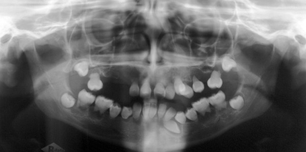
Background: Dentin dysplasia type I is characterized by a defect of dentin development with clinical normal appearance of the permanent teeth but no or only rudimentary root formation. Early loss of all teeth and concomitant underdevelopment of the jaws are challenging for successful treatment with dental implants.

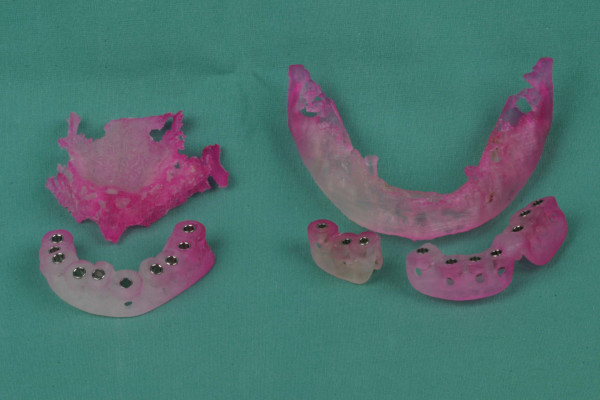
Methods: A combination of sinus lifting and onlay bone augmentation based on treatment planning using stereolithographic templates was used in a patient with dentin dysplasia type I to rehabilitate the masticatory function.

Results: (i) a predisposition for an increased and accelerated bone resorption was observed in our patient, (ii) bone augmentation was successful using a mixture of allogenic graft material with autogenous bone preventing fast bone resorption, (iii) surgical planning, based on stereolithographic models and surgical templates, facilitated the accurate placement of dental implants.

Conclusion: Bony augmentation and elaborate treatment planning is helpful for oral rehabilitation of patients with dentin dysplasia type I.


Figures

Figure 1
Figure 1
initial clinical situation.
Figure 2
Figure 2
preoperative panoramic radiographs showing features of dentin dysplasia type I.
Figure 3
Figure 3
alveolar ridge augmentation of the maxilla (above) and the mandible (below) using autogenous bone grafts from the iliac crest.
Figure 4
Figure 4
postoperative panoramic radiographs after tooth extraction and bone augmentation.
Figure 5
Figure 5
stereolithographic templates with drill-guide tubes manufactured on three-dimensional stereolithographic models of the mandible and maxilla.
Figure 6
Figure 6
postoperative panoramic radiographs after implant setting and bone augmentation.
Figure 7
Figure 7
postoperative clinical situation after completion of the implant treatment.

References

    1. O Carroll MK, Duncan WK. Dentin dysplasia type I. Radiologic and genetic perspectives in a six-generation family. Oral Surg Oral Med Oral Pathol. 1994;78:375–381. doi: 10.1016/0030-4220(94)90071-X. - DOI - PubMed
    1. Shields ED, Bixler D, el-Kafrawy AM. A proposed classification for heritable human dentine defects with a description of a new entity. Arch Oral Biol. 1973;18:543–553. doi: 10.1016/0003-9969(73)90075-7. - DOI - PubMed
    1. O Carroll MK, Duncan WK, Perkins TM. Dentin dysplasia: review of the literature and a proposed subclassification based on radiographic findings. Oral Surg Oral Med Oral Pathol. 1991;72:119–125. doi: 10.1016/0030-4220(91)90202-N. - DOI - PubMed
    1. Ommerborn M, Raab W. Allgemeinerkrankungen und Schäden der Zahnhartsubstanzen. Prophylaxe Impuls. 2005;9:66–73.
    1. Pettiette MT, Wright JT, Trope M. Dentinogenesis imperfecta: endodontic implications. Case report. Oral Surg Oral Med Oral Pathol Oral Radiol Endod. 1998;86:733–737. doi: 10.1016/S1079-2104(98)90213-X. - DOI - PubMed

LinkOut - more resources