

Site-directed in vitro immunization leads to a complete human monoclonal IgG4 lambda that binds specifically to the CDR2 region of CTLA-4 (CD152) without interfering the engagement of natural ligands
- PMID: 17714596
- PMCID: PMC2025598
- DOI: 10.1186/1472-6750-7-51
Site-directed in vitro immunization leads to a complete human monoclonal IgG4 lambda that binds specifically to the CDR2 region of CTLA-4 (CD152) without interfering the engagement of natural ligands
Abstract
Background: The ability to acquire fully human monoclonal antibodies (mAbs) with pre-defined specificities is critical to the development of molecular tags for the analysis of receptor function in addition to promising immunotherapeutics. Yet most of the arriving affinity maturated and complete human immunoglobulin G (IgG) molecules, which are actually derived from single human B cells, have not widely been used to study the conserved self antigens (Ags) such as CD152 (cytotoxic T lymphocyte antigen-4, CTLA-4) because proper hosts are lacking.
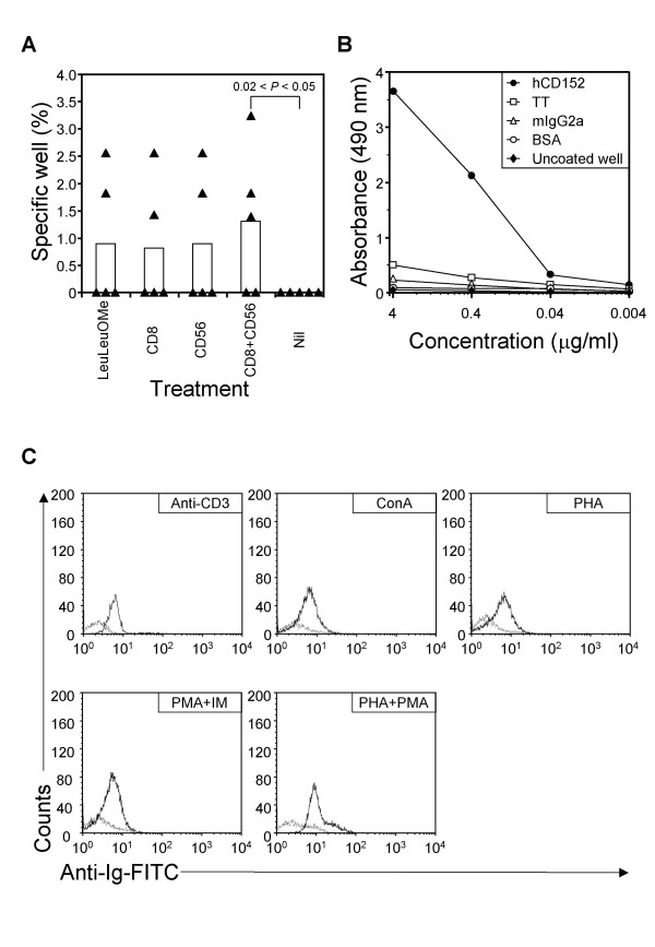
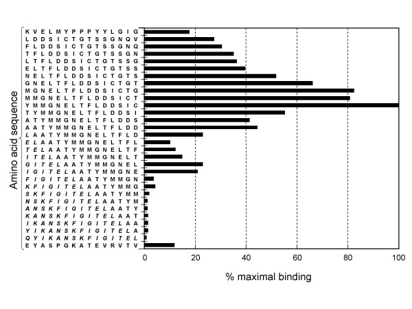
Results: Here we developed an optimized protocol for site-directed in vitro immunizing peripheral blood mononuclear cells (PBMC) by using a selected epitope of human CD152, an essential receptor involved in down-regulation of T cell activation. The resultant stable trioma cell lines constantly produce anti-CD152 mAb (gamma4lambdahuCD152), which contains variable (V) regions of the heavy chain and the light chain derived from the VH3 and V lambda human germline genes, respectively, and yet displays an unusual IgG4 isotype. Interestingly, gamma4lambdahuCD152 has a basic pI not commonly found in myeloid monoclonal IgG4 lambdas as revealed by the isoelectric focusing (IEF) analysis. Furthermore, gamma4lambdahuCD152 binds specifically, with nanomolar affinity, to an extracellular constituency encompassing the putative second complementarity determining region (CDR2) of CD152, whereby it can react to activated CD3+ cells.
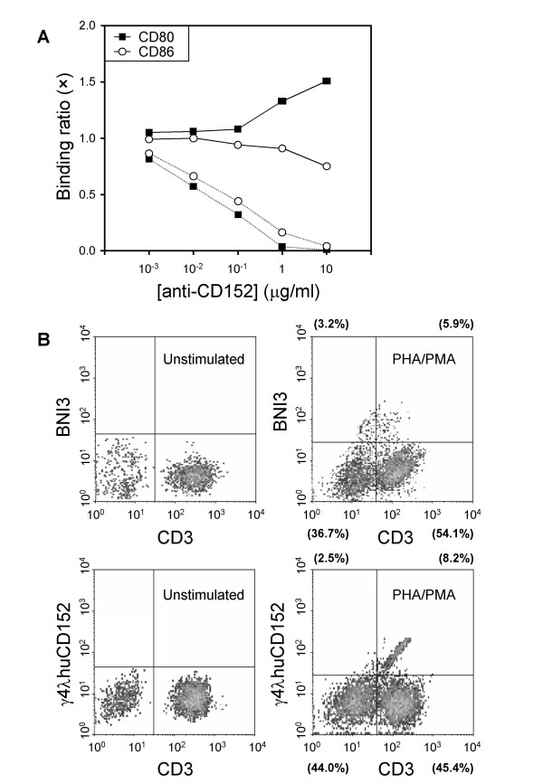
Conclusion: In a context of specific cell depletion and conditioned medium,in vitro induction of human Abs against a conserved self Ag was successfully acquired and a relatively basic mAb, gamma4lambdahuCD152, with high affinity to CDR2 of CD152 was thus obtained. Application of such a human IgG4 lambda mAb with designated CDR2 specificity may impact upon and prefer for CD152 labeling both in situ and ex situ, as it does not affect the binding of endogenous B7 ligands and can localize into the confined immunological synapse which may otherwise prevent the access of whole IgG1 molecules.
Figures


Similar articles
-
Building novel binding ligands to B7.1 and B7.2 based on human antibody single variable light chain domains.J Mol Biol. 2001 Jul 13;310(3):591-601. doi: 10.1006/jmbi.2001.4703. J Mol Biol. 2001. PMID: 11439026
-
Functional humanization of an anti-CD16 Fab fragment: obstacles of switching from murine {lambda} to human {lambda} or {kappa} light chains.Protein Eng Des Sel. 2009 Mar;22(3):175-88. doi: 10.1093/protein/gzn066. Epub 2008 Nov 19. Protein Eng Des Sel. 2009. PMID: 19022801
-
Amplification of immunoglobulin transcripts by the non-palindromic adaptor polymerase chain reaction (NPA-PCR). Nucleotide sequence analysis of two human monoclonal antibodies recognizing two cell surface antigens expressed in ovarian, cervix, breast, colon and other carcinomas.Hum Antibodies Hybridomas. 1994;5(3-4):131-42. Hum Antibodies Hybridomas. 1994. PMID: 7756578
-
Immune intervention with monoclonal antibodies targeting CD152 (CTLA-4) for autoimmune and malignant diseases.Chang Gung Med J. 2008 Jan-Feb;31(1):1-15. Chang Gung Med J. 2008. PMID: 18419049 Review.
-
Recombinant antibodies.Hum Antibodies Hybridomas. 1991 Oct;2(4):172-89. Hum Antibodies Hybridomas. 1991. PMID: 1751781 Review.
Cited by
-
Production of antigen-specific human IgGs by in vitro immunization.BMC Biotechnol. 2016 Feb 24;16:22. doi: 10.1186/s12896-016-0253-1. BMC Biotechnol. 2016. PMID: 26911296 Free PMC article.
-
Long-term immunologically competent human peripheral lymphoid tissue cultures in a 3D bioreactor.Biotechnol Bioeng. 2011 Jun;108(6):1430-40. doi: 10.1002/bit.23055. Epub 2011 Feb 18. Biotechnol Bioeng. 2011. PMID: 21309085 Free PMC article.
References
-
- Magadán S, Valladares M, Suarez E, Sanjuán I, Molina A, Ayling C, Davies SL, Zou X, Williams GT, Neuberger MS, Brüggemann M, Gambón F, Diíz-Espada F, González-Fernández A. Production of antigen-specific human monoclonal antibodies: comparison of mice carrying IgH/kappa or IgH/kappa/lambda transloci. Biotechniques. 2002;33:680–690. - PubMed
MeSH terms
Substances
Associated data
- Actions
- Actions
LinkOut - more resources
Full Text Sources
Other Literature Sources
Research Materials
Miscellaneous
