. 2007 Oct;207(2):186-94.
doi: 10.1016/j.expneurol.2007.06.028. Epub 2007 Jul 26.

Slit modulates cerebrovascular inflammation and mediates neuroprotection against global cerebral ischemia

Affiliations

Slit modulates cerebrovascular inflammation and mediates neuroprotection against global cerebral ischemia

Tamer Altay et al. Exp Neurol. 2007 Oct.

Abstract

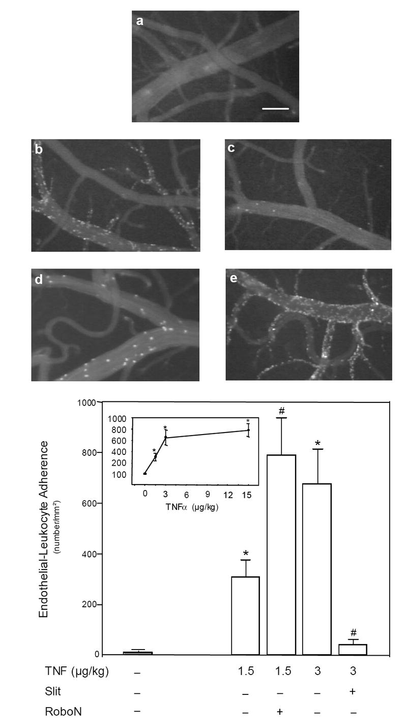
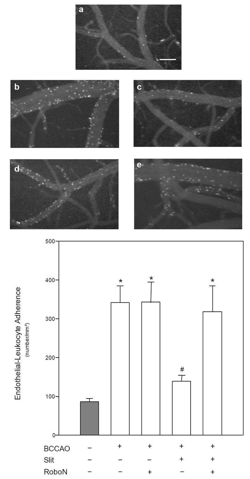
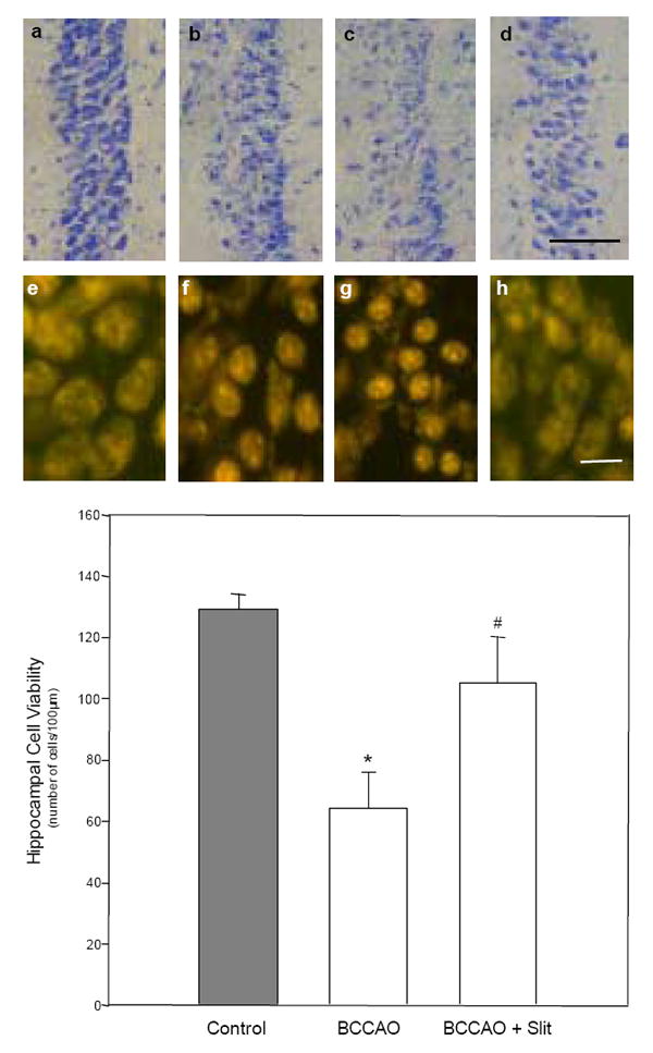
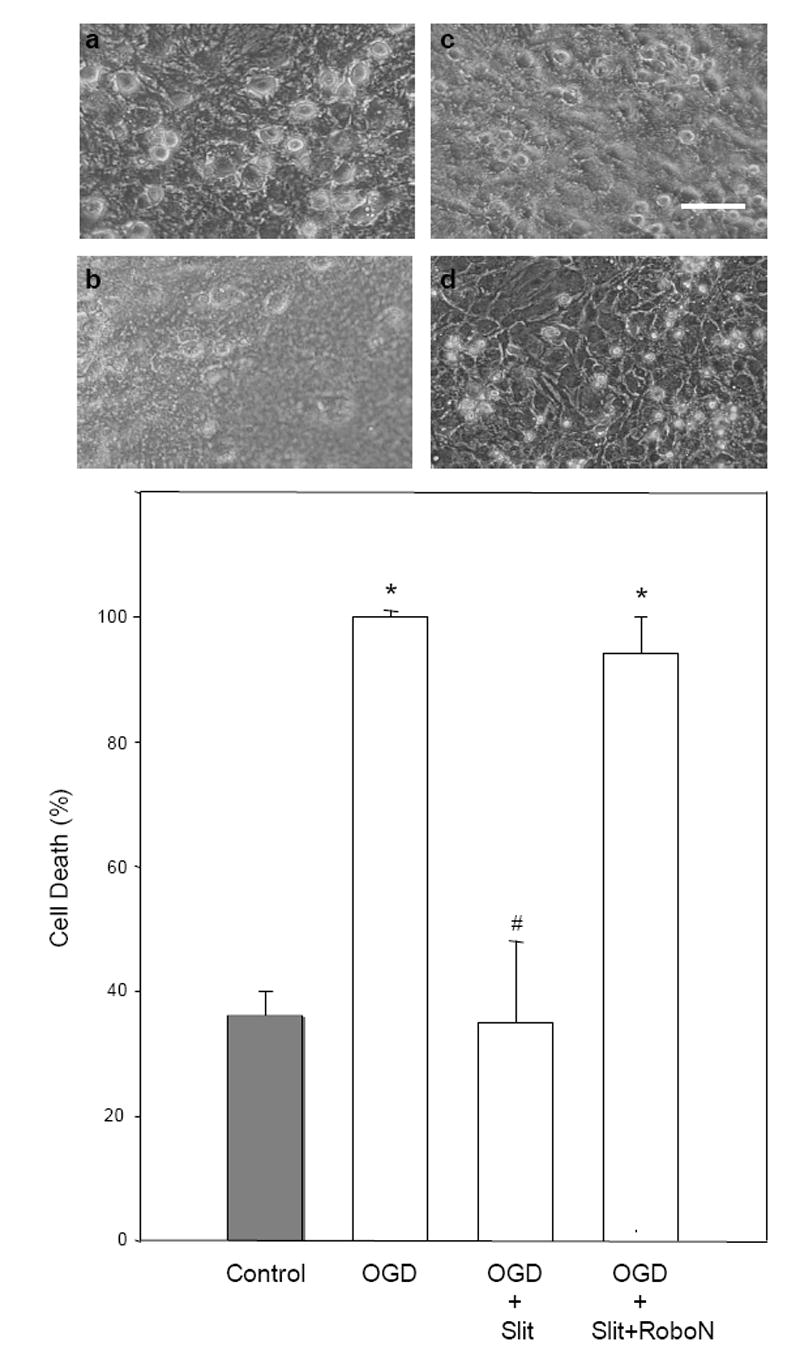
Cerebrovascular inflammation contributes to secondary brain injury following ischemia. Recent in vitro studies of cell migration and molecular guidance mechanisms have indicated that the Slit family of secreted proteins can exert repellant effects on leukocyte recruitment in response to chemoattractants. Utilizing intravital microscopy, we addressed the role of Slit in modulating leukocyte dynamics in the mouse cortical venular microcirculation in vivo following TNFalpha application or global cerebral ischemia. We also studied whether Slit affected neuronal survival in the mouse global ischemia model as well as in mixed neuronal-glial cultures subjected to oxygen-glucose deprivation. We found that systemically administered Slit significantly attenuated cerebral microvessel leukocyte-endothelial adherence occurring 4 h after TNFalpha and 24 h after global cerebral ischemia. Administration of RoboN, the soluble receptor for Slit, exacerbated the acute chemotactic response to TNFalpha. These findings are indicative of a tonic repellant effect of endogenous Slit in brain under acute proinflammatory conditions. Three days of continuous systemic administration of Slit following global ischemia significantly attenuated the delayed neuronal death of hippocampal CA1 pyramidal cells. Moreover, Slit abrogated neuronal death in mixed neuronal-glial cultures exposed to oxygen-glucose deprivation. The ability of Slit to reduce the recruitment of immune cells to ischemic brain and to provide cytoprotective effects suggests that this protein may serve as a novel anti-inflammatory and neuroprotective target for stroke therapy.


Figures

Figure 1
Figure 1
Effect of Slit on TNFα-induced increases in leukocyte-endothelial adherence. Representative epifluorescent videophotomicrographs of cortical venular leukocytes in the different groups are shown, relative to untreated controls (a). TNFα (3 μg/kg) induced leukocyte adherence to cerebral venular endothelium (b) and this response was completely blocked by exogenous Slit (c). The increase in leukocyte-endothelial adherence induced by TNFα (1.5 μg/kg) (d) was exacerbated by the soluble receptor RoboN (e), reflecting tonic inhibition of adherence by endogenous Slit. Histogram summarizes the magnitude of leukocyte-endothelial adherence under the different experimental conditions, relative to naive controls (shaded bar). Inset: Dose dependency of TNFα-induced inflammation. Intravital imaging was performed 4 h after animals were administered TNFα. *p<0.05 vs. sham; #p<0.05 vs. TNFα only (at the corresponding dose). Scale bar = 100μm.
Figure 2
Figure 2
Effect of Slit on increases in leukocyte-endothelial adherence induced by global ischemia (bilateral common carotid occlusion [BCCAO]). Representative epifluorescent videophotomicrographs of cortical venular leukocytes in the different groups are shown, relative to sham BCCAO controls (a). BCCAO-induced leukocyte adherence to cerebral venular endothelium (b) was blocked by co-administration of Slit (c). The soluble receptor RoboN blocked Slit’s inhibitory effect on BCCAO-induced adherence (d) but did not exacerbate BCCAO-induced adherence (e). Histogram summarizes the magnitude of leukocyte-endothelial adherence under the different experimental conditions, relative to naive controls (shaded bar). Animals received 6 min BCCAO and recovered 24 h before intravital imaging of leukocyte-endothelial adherence. *p<0.05 vs. sham; #p<0.05 vs. BCCAO. Scale bar = 100μm.
Figure 3
Figure 3
Effect of Slit on CA1 pyramidal cell injury following global ischemia. Representative Nissl-stained (a-d, first row) and propidium iodide-stained (e-h, second row) hippocampal CA1 pyramidal cells 7 days after global ischemia (bilateral common carotid occlusion [BCCAO]). Relative to naïve controls (a,e), in animals with both moderate (~50% cell loss; b,f) and severe (~80% cell loss; c,g) CA1 injury, PI-labeled cells displayed distinct, multiple, hyperintense nuclear condensations. However, in animals treated with Slit (d,h), a more viable pyramidal cell morphology was the prominent feature. The histogram quantifies the protective effect of exogenous Slit on hippocampal CA1 pyramidal cell viability, relative to naive controls (shaded bar). *p<0.05 vs. control; #p<0.05 vs. BCCAO. Scale bar = 50μm and 10μm in Nissl- and PI-stained sections, respectively.
Figure 4
Figure 4
Effects of Slit on oxygen-glucose deprivation (OGD)/reoxygenation injury in mixed neuronal-glial cultures. Representative photomicrographs are provided for each experimental condition. (a) Under untreated control conditions, neurons were healthy, phase bright, and had well defined processes. (b) 90-min OGD resulted in robust neuronal cell death with marked cellular debris evident the following day. (c) Slit significantly and dramatically attenuated neuronal cell death induced by 90-min OGD and recovery. Although some morphological changes occurred, including slight decreases in cell volume, neurons remained viable (as assessed by LDH assay). (d) The protective effect of Slit was reversed by co-incubation with its soluble receptor RoboN. Histogram shows quantification of neuronal cell death as assessed by release of lactate dehydrogenase (LDH) into the media, normalized to total kill (100%), relative to naive, untreated controls (shaded bar) over the same time period. *p<0.001 vs. Control; #p<0.001 vs. OGD. Scale bar = 40μm

References

    1. Adamson P, Etienne S, Couraud PO, Calder V, Greenwood J. Lymphocyte migration through brain endothelial cell monolayers involves signaling through endothelial ICAM-1 via a rho-dependent pathway. J Immunol. 1999;162:2964–2973. - PubMed
    1. Akopov SE, Simonian NA, Grigorian GS. Dynamics of polymorphonuclear leukocyte accumulation in acute cerebral infarction and their correlation with brain tissue damage. Stroke. 1996;27:1739–1743. - PubMed
    1. Alon R, Grabovsky V, Feigelson S. Chemokine induction of integrin adhesiveness on rolling and arrested leukocytes local signaling events or global stepwise activation? Microcirculation. 2003;10:297–311. - PubMed
    1. Altay T, Gonzales ER, Park TS, Gidday JM. Cerebrovascular inflammation after brief episodic hypoxia: modulation by neuronal and endothelial nitric oxide synthase. J Appl Physiol. 2004;96:1223–1230. - PubMed
    1. Aspey BS, Jessimer C, Pereira S, Harrison MJ. Do leukocytes have a role in the cerebral no-reflow phenomenon? J Neurol Neurosurg Psychiatr. 1989;52:526–528. - PMC - PubMed

Publication types

MeSH terms