. 2007 Aug 28;104(35):13942-7.
doi: 10.1073/pnas.0706404104. Epub 2007 Aug 21.

Crystal structure of the incretin-bound extracellular domain of a G protein-coupled receptor

Affiliations

Crystal structure of the incretin-bound extracellular domain of a G protein-coupled receptor

Christoph Parthier et al. Proc Natl Acad Sci U S A. .

Abstract

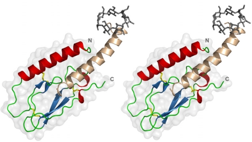
Incretins, endogenous polypeptide hormones released in response to food intake, potentiate insulin secretion from pancreatic beta cells after oral glucose ingestion (the incretin effect). This response is signaled by the two peptide hormones glucose-dependent insulinotropic polypeptide (GIP) (also known as gastric inhibitory polypeptide) and glucagon-like peptide 1 through binding and activation of their cognate class 2 G protein-coupled receptors (GPCRs). Because the incretin effect is lost or significantly reduced in patients with type 2 diabetes mellitus, glucagon-like peptide 1 and GIP have attracted considerable attention for their potential in antidiabetic therapy. A paucity of structural information precludes a detailed understanding of the processes of hormone binding and receptor activation, hampering efforts to develop novel pharmaceuticals. Here we report the crystal structure of the complex of human GIP receptor extracellular domain (ECD) with its agonist, the incretin GIP(1-42). The hormone binds in an alpha-helical conformation in a surface groove of the ECD largely through hydrophobic interactions. The N-terminal ligand residues would remain free to interact with other parts of the receptor. Thermodynamic data suggest that binding is concomitant with structural organization of the hormone, resulting in a complex mode of receptor-ligand recognition. The presentation of a well structured, alpha-helical ligand by the ECD is expected to be conserved among other hormone receptors of this class.


Conflict of interest statement

The authors declare no conflict of interest.

Figures

Fig. 1.
Fig. 1.
Overall structure of the GIPR ECD in complex with GIP1–42. Stereoview of the peptide hormone GIP1–42 (residues 1′–32′ beige; residues 33′–42′ were not visible in the electron density) bound to the ECD of GIPR [colored according to secondary structure, residues 29(N)–122(C) visible]. Disulfide bridges in the GIPR ECD are shown as yellow sticks. The N terminus of the ligand is bound by a methyl-β-cyclodextrin molecule (gray sticks).
Fig. 2.
Fig. 2.
Hormone receptor ECDs and ligands. (a) Sequence alignment of the ECDs of human hormone receptors (CRFR-2β: murine sequence). The numbering is according to GIPR residues, and the names of the glucagon receptor family members are in bold. The conserved cysteines are marked in yellow, other absolutely conserved residues are in red, and less conserved amino acids are highlighted in gray (hydrophobic) and green (basic), respectively. The dots in the sequence represent additional residues that are not shown. Amino acids of the predicted N-terminal α-helix are underlined; residues missing from the construct of CRFR-2β used for structure determination (26) are in gray. Amino acids involved in stabilizing the GIPR ECD core structure are marked by asterisks in the line “fold.” (b) Sequence alignment of human peptide hormones (exendin-4 from Heloderma suspectum), with numbering according to GIP; the names of glucagon receptor family ligands are in bold. The absolutely conserved Phe-6′ is marked in red, and the positions of hydrophobic amino acids corresponding to the GIP residues interacting with the ECD are highlighted in gray. Potential N-terminal helix-capping residues are marked in green, and PACAP residues forming an α-helix when bound to its receptor are underlined (47). The sequence of astressin used in the NMR structure determination (26) is aligned according to superposition of the ECDs of GIPR and CRFR-2β; f denotes DPhe12, m indicates norleucine residues 21 and 38, and underlined residues Glu-30 and Lys-33 are chemically linked through a lactam bridge. Residues not observed in the NMR structure are in gray, and those involved in ECD binding are highlighted. The GIPR ECD and GIP1–42 residues comprising the binding interface are marked with asterisks in the line “binding”; red asterisks are assigned to residues involved in hydrophobic interactions. The secondary structure found in the structure of the complex is depicted in the line “ss.”
Fig. 3.
Fig. 3.
The glucagon hormone family recognition fold. (a) Stereoview of the GIPR ECD with the three clusters of residues characteristic for the domain structure, consisting of the following: W71, W109, R101, and V99 (cluster 1, magenta); residues W39, Y42, and F65 (cluster 2, dark blue); and residues P85, Y68, Y87, L88, and W90 (cluster 3, cyan). The absolutely conserved D66 is shown in green, and disulfide bridges are in yellow. (b) Stick representation of the GIPR ECD loop region around D66 (green), with a 2FoFc map at 1.5σ contour (blue mesh), and of GIP residues L26′ and L27′ (orange), with corresponding 2FoFc map (gray mesh). The side chain of D66 is involved in hydrogen bonds (dashed lines) to W71 (magenta) and residues M67, Y67, and V69 (shown in cyan) and thereby fixes the loop that is involved in ligand binding.
Fig. 4.
Fig. 4.
Hydrophobic interactions involved in ligand binding. Stereoview of GIP1–42 (beige) bound to the GIPR ECD (white) with key ligand residues (stick representation, orange) that occupy the hydrophobic binding groove of the GIPR ECD formed by residues from cluster 2 (dark blue), cluster 3 (cyan), the D66-loop region (magenta), or other residues at the ECD surface (green).

References

    1. Fehmann HC, Göke R, Göke B. Endocr Rev. 1995;16:390–410. - PubMed
    1. Drucker DJ. Cell Metab. 2006;3:153–165. - PubMed
    1. Nauck MA, Baller B, Meier JJ. Diabetes. 2004;53(Suppl 3):S190–S196. - PubMed
    1. O'Harte FP, Gray AM, Flatt PR. J Endocrinol. 1998;156:237–243. - PubMed
    1. Xie D, Cheng H, Hamrick M, Zhong Q, Ding KH, Correa D, Williams S, Mulloy A, Bollag W, Bollag RJ, et al. Bone. 2005;37:759–769. - PubMed

Publication types

Associated data