. 2007 Nov;81(21):11861-9.
doi: 10.1128/JVI.01077-07. Epub 2007 Aug 22.

Three-dimensional structure of the human cytomegalovirus cytoplasmic virion assembly complex includes a reoriented secretory apparatus

Affiliations

Three-dimensional structure of the human cytomegalovirus cytoplasmic virion assembly complex includes a reoriented secretory apparatus

Subhendu Das et al. J Virol. 2007 Nov.

Abstract

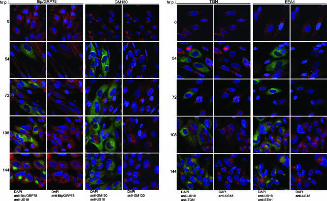
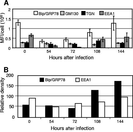
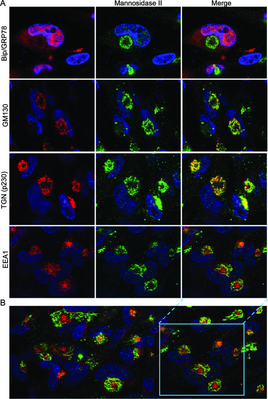
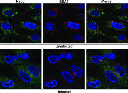
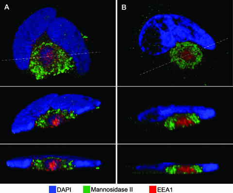
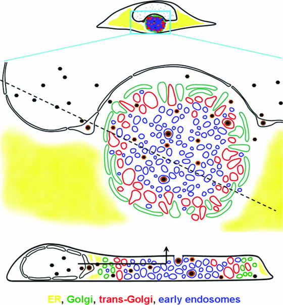
Human cytomegalovirus (HCMV) induces profound changes in infected cell morphology, including a large cytoplasmic inclusion that corresponds to the virion assembly complex (AC). In electron micrographs, the AC is a highly vacuolated part of the cytoplasm. Markers of cellular secretory organelles have been visualized at the outer edge of the AC, and we recently showed that a marker for early endosomes (i.e., early endosome antigen 1) localizes to the center of the AC. Here, we examined the relationship between the AC and components of the secretory apparatus, studied temporal aspects of the dramatic infection-induced cytoplasmic remodeling, examined the three-dimensional structure of the AC, and considered the implications of our observations for models of HCMV virion maturation and egress. We made three major observations. First, in addition to being relocated, the expression levels of some organelle markers change markedly during the period while the AC is developing. Second, based on three-dimensional reconstructions from z-series confocal microscopic images, the observed concentric rings of vesicles derived from the several compartments (Golgi bodies, the trans-Golgi network [TGN], and early endosomes) are arranged as nested cylinders of organelle-specific vesicles. Third, the membrane protein biosynthetic and exocytic pathways from the endoplasmic reticulum to the Golgi bodies, TGN, and early endosomes are in an unusual arrangement that nonetheless allows for a conventional order of biosynthesis and transport. Our model of AC structure suggests a mechanism by which the virus can regulate the order of tegument assembly.


Figures

FIG. 1.
FIG. 1.
Time course of cytoplasmic remodeling. HLF cells were mock infected or infected for 144 h at an MOI of 0.01. The indicated organelle markers (ER, Bip/GRP78; Golgi, GM130; TGN, p230; and early endosomes, EEA1) were detected by confocal microscopy using MAb (red stain), and infected cells were detected with a rabbit polyclonal antibody to US18 (green). DNA was stained with DAPI. For each organelle and time point, the left panel is a merge of all three colors, and the right panel is a merge of just the green and blue channels.
FIG. 2.
FIG. 2.
Time course of expression of components of the secretory apparatus in HCMV-infected cells. (A) Images collected in the course of experiments similar to those shown in Fig. 1 were analyzed to quantitate the mean fluorescence intensity (MFI) in a single confocal cross-section per cell for each marker. Ten infected cells were analyzed for each marker. (B) Relative immune blot reactivity of Bip/GRP78 and EEA1 as a function of time after infection, as measured by densitometry of chemiluminescence images. Organelle markers: ER, Bip/GRP78; Golgi, GM130; TGN, p230; and early endosomes, EEA1.
FIG. 3.
FIG. 3.
Localization of secretory apparatus components relative to a Golgi marker. (A) HLF cells mock infected or infected with HCMV(AD169) for 144 h were fixed and then stained with a rabbit polyclonal antibody against a Golgi marker (mannosidase II) (green), MAb against the indicated organelle markers (red), and DAPI prior to confocal microscopy. The organelle markers detected with the MAb were Bip/GRP78 (ER), GM130 (Golgi), p230 (TGN), and EEA1 (early endosomes). (B) A larger field from the early endosome/Golgi staining is shown to demonstrate that the patterns shown are representative.
FIG. 4.
FIG. 4.
Relative localization of the early endosome-associated proteins, EEA1 and rab5. HLF cells were infected with HCMV(AD169) for 144 h before being fixed and stained with DAPI (blue), a mouse MAb against the early endosomal marker EEA1 (red), and a rabbit polyclonal antibody the rab5 (green). The images were obtained by confocal microscopy.
FIG. 5.
FIG. 5.
Three-dimensional reconstructions of HCMV-infected cell structure. HLF cells were infected with HCMV(AD169) for 108 h (A) or 144 h (B and C) before being fixed and stained with DAPI (blue), a mouse MAb against the early endosomal marker EEA1 (red), and a rabbit polyclonal antibody the Golgi marker mannosidase II (green). A z-series of confocal microscopic images was obtained at 1-μm increments. In each panel, the top image is a projection of the complete z-series. The middle images are tilted cross-sections taken along the dotted line in panel A. The dotted boxes outline the cut face. The bottom images in each panel are edge-on views of the same section (perpendicular to the z-axis). Note that the green Golgi marker does not extend across the top of the cut face, which is evidence for a nested cylindrical, rather than nested spherical, arrangement of organelle-specific vesicles.
FIG. 6.
FIG. 6.
Schematic representation of AC structure and the maturational path of nascent virions. The diagram is based on data such as shown in Fig. 1, 3, and 5, coupled with prior ultrastructural analyses (see, for example, reference 50) and other information cited in the text about herpesvirus maturation. In this representation, the AC is the large circular structure that is bounded by Golgi vesicles. The concentric arrangement of the Golgi, TGN, and early endosomal compartments is shown. The cross-sectional representation through the cell at the bottom of the figure shows the nested cylindrical arrangement of the vesicular compartments (sectioning along the dotted line in the upper panel). In this model, nascent capsids acquire a subset of tegument proteins prior to nuclear egress. In the cytoplasm, tegumentation occurs during migration from the AC periphery to the exit vesicle, which is transported vertically to the cell surface without needing to traverse the secretory pathway in reverse. The path of egress is indicated in the lower panel by the arrow.

Similar articles

Cited by

References

    1. AuCoin, D. P., G. B. Smith, C. D. Meiering, and E. S. Mocarski. 2006. Betaherpesvirus-conserved cytomegalovirus tegument protein ppUL32 (pp150) controls cytoplasmic events during virion maturation. J. Virol. 80:8199-8210. - PMC - PubMed
    1. Azzeh, M., A. Honigman, A. Taraboulos, A. Rouvinski, and D. G. Wolf. 2006. Structural changes in human cytomegalovirus cytoplasmic assembly sites in the absence of UL97 kinase activity. Virology 354:69-79. - PubMed
    1. Barr, F. A. 1999. A novel Rab6-interacting domain defines a family of Golgi-targeted coiled-coil proteins. Curr. Biol. 9:381-384. - PubMed
    1. Barr, F. A., and B. Short. 2003. Golgins in the structure and dynamics of the Golgi apparatus. Curr. Opin. Cell Biol. 15:405-413. - PubMed
    1. Benboudjema, L., M. Mulvey, Y. Gao, S. W. Pimplikar, and I. Mohr. 2003. Association of the herpes simplex virus type 1 Us11 gene product with the cellular kinesin light-chain-related protein PAT1 results in the redistribution of both polypeptides. J. Virol. 77:9192-9203. - PMC - PubMed