. 2007 Aug 22;27(34):9032-42.
doi: 10.1523/JNEUROSCI.2088-07.2007.

Angiotensin II controls occludin function and is required for blood brain barrier maintenance: relevance to multiple sclerosis

Affiliations

Angiotensin II controls occludin function and is required for blood brain barrier maintenance: relevance to multiple sclerosis

Karolina Wosik et al. J Neurosci. .

Abstract

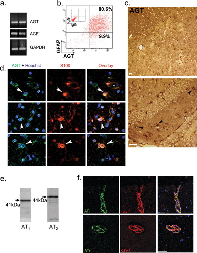
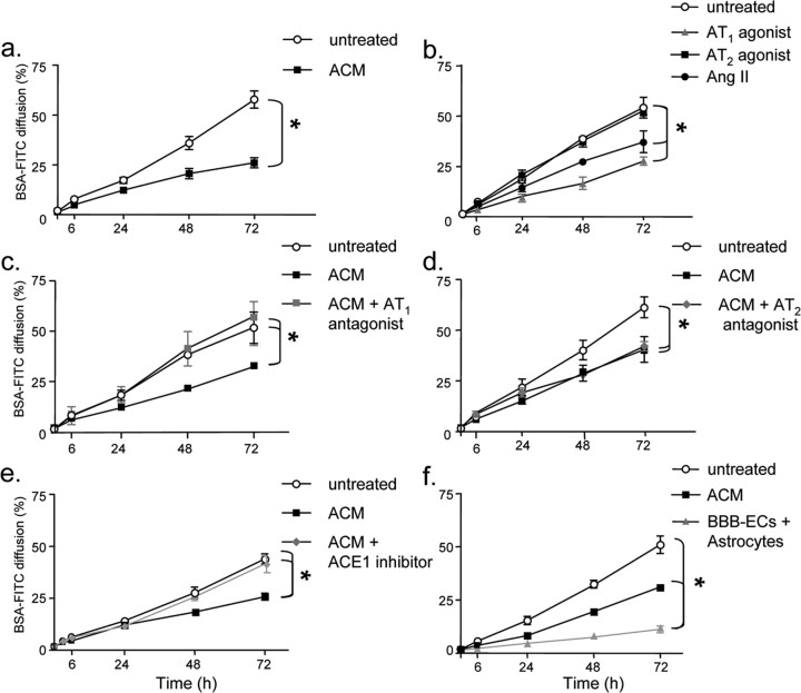
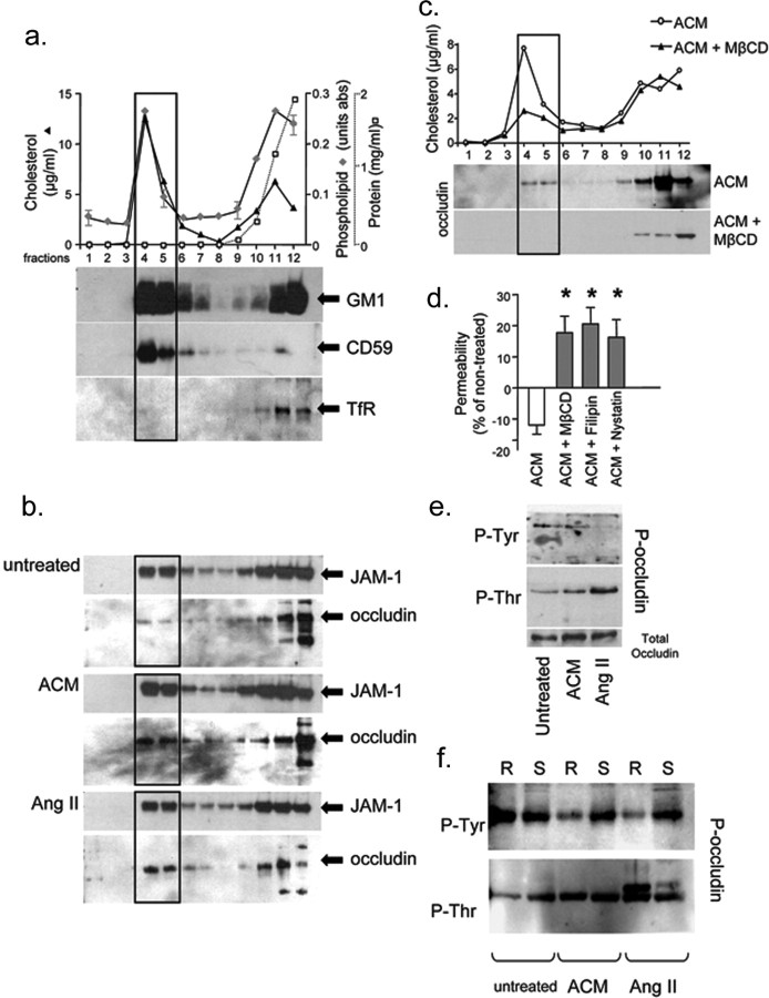
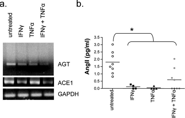
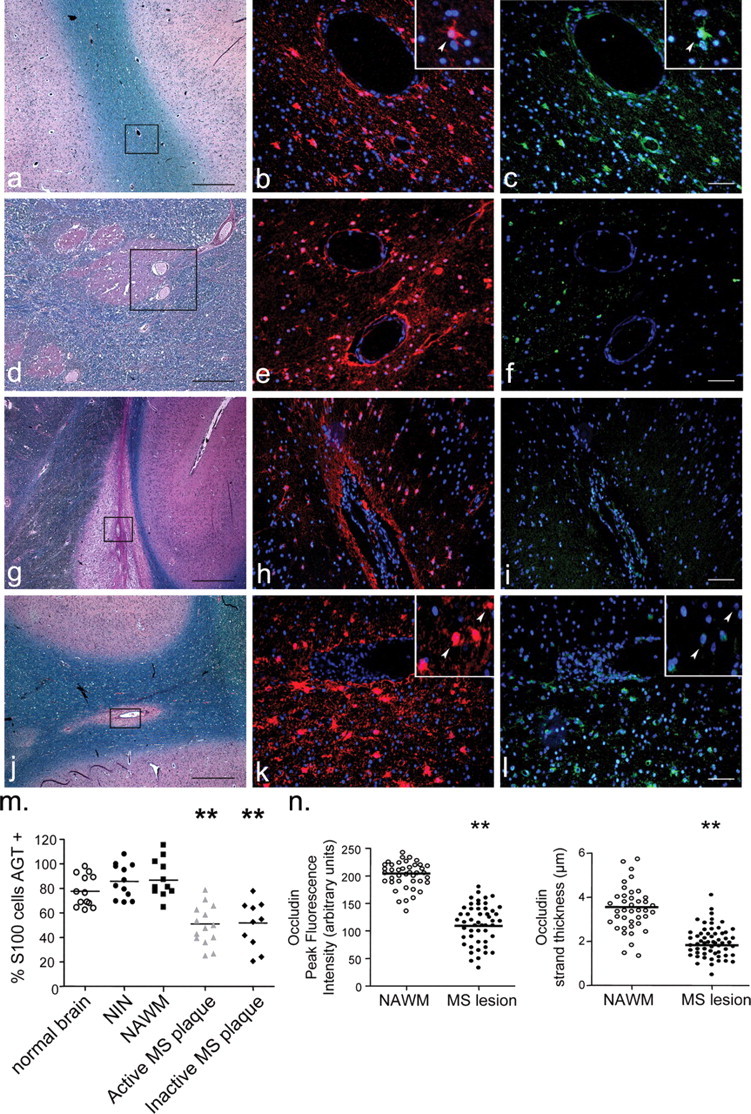
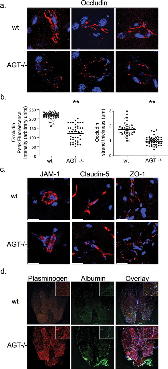
The blood-brain barrier (BBB) restricts molecular and cellular trafficking between the blood and the CNS. Although astrocytes are known to control BBB permeability, the molecular determinants of this effect remain unknown. We show that angiotensinogen (AGT) produced and secreted by astrocytes is cleaved into angiotensin II (AngII) and acts on type 1 angiotensin receptors (AT1) expressed by BBB endothelial cells (ECs). Activation of AT1 restricts the passage of molecular tracers across human BBB-derived ECs through threonine-phosphorylation of the tight junction protein occludin and its mobilization to lipid raft membrane microdomains. We also show that AGT knock-out animals have disorganized occludin strands at the level of the BBB and a diffuse accumulation of the endogenous serum protein plasminogen in the CNS, compared with wild-type animals. Finally, we demonstrate a reduction in the number of AGT-immunopositive perivascular astrocytes in multiple sclerosis (MS) lesions, which correlates with a reduced expression of occludin similarly seen in the CNS of AGT knock-out animals. Such a reduction in astrocyte-expressed AGT and AngII is dependent, in vitro, on the proinflammatory cytokines tumor necrosis factor-alpha and interferon-gamma. Our study defines a novel physiological role for AngII in the CNS and suggests that inflammation-induced downregulation of AngII production by astrocytes is involved in BBB dysfunction in MS lesions.


Figures

Figure 1.
Figure 1.
Angiotensinogen, Angiotensin II, and angiotensin receptor expression in human brain. a, PCR of human astrocytes in culture for AGT and ACE1, two astrocyte preparations are shown. b, Flow cytometry analysis of human fetal astrocytes in culture immunostained for GFAP and AGT (n = 3). c, Immunohistochemistry on normal human CNS sections (hemispheric and cerebellar) reveals the presence of numerous AGT-positive cells (n = 8 sections; 2 CNS samples). Scale bars, 20 μm. d, Immunohistofluorescence on normal human subcortical white matter shows S100-positive astrocytes (red) are immunopositive for AGT (green), as evidenced by the yellow color in the overlay pictures. Vessel lumen is shown with an asterisk. Nuclei were stained with Hoechst (blue). e, Western blots for angiotensin receptors AT1 and AT2 on human BBB-ECs. f, Immunohistofluorescence of normal human subcortical white matter shows brain capillaries are immunopositive for both cav-1 (red) as well as for AT1 and AT2 (green); overlays are shown on the right; TO-PRO3-stained nuclei are blue. Scale bars, 50 μm.
Figure 2.
Figure 2.
Astrocyte-secreted angiotensin II decreases endothelial cell permeability via the AT1 receptor. a, Permeability of BBB-EC monolayers grown in the modified Boyden chamber assay to FITC-BSA is substantially decreased when cells are grown in the presence of astrocyte-conditioned media (filled square; ACM 40% v/v) when compared with cells grown under basal conditions (open circle; untreated) (n = 3, in duplicate; *p < 0.005, p < 0.0001, Bonferroni posttest at 48 and 72 h, respectively). b, A similar decrease in permeability is seen when BBB-ECs are grown in the presence of AngII (filled circle;10−8 m, AngII) or the angiotensin receptor AT1 agonist L-162,313 (filled triangle; 10−8 m; n = 3, in duplicate; *p < 0.005, compared with nontreated cells). AT2 agonist CGP-42112A (filled square; 10−8 m) did not affect permeability. c, d, BBB-EC monolayers were grown in the presence of ACM with or without the AT1 receptor antagonist Losartan (c) (filled square; 10−7 m) or the AT2 receptor antagonist PD123,319 (d) (filled diamond; 10−7 m). The AT1 receptor antagonist blocks the permeability decreasing the effect of ACM; the AT2 antagonist does not (n = 3; in duplicate, *p < 0.005 between indicated curves). e, Astrocyte cultures were treated with ACE1 inhibitor (filled diamond; Captopril, 5 d at 10−7 m) to reduce AngII levels in ACM. Captopril-treated ACM did not promote BBB integrity, compared with untreated ACM (n = 3, in duplicate; *p < 0.005 between indicated curves). f, BBB-ECs were cocultured with astrocytes (filled triangle), with ACM (filled square), or alone (open circle) for 72 h to demonstrate that astrocyte-EC proximity is important for barrier properties (n = 2, in duplicate; *p < 0.01). All data are expressed as mean ± SEM.
Figure 3.
Figure 3.
Lipid raft profiles of blood–brain barrier endothelial cells. a, Characterization of BBB-EC sucrose density fractions with regards to cholesterol concentration (filled triangle; left axis; in μg/ml), phospholipid content (filled diamond; right axis; in values of absorbance), and protein concentration (open square; far right axis; in mg/ml). Lipid rafts are concentrated in fractions 4 and 5 (rectangular outline) where peak values of cholesterol and phospholipids are seen. Fractions 4 and 5 also show a high concentration of lipid raft markers GM1 ganglioside and CD59 but no evidence of transferrin receptor (TfR), a marker of nonlipid raft membrane. b, Western blots for the TJ protein JAM-1 in sucrose density fractions show a similar concentration of JAM-1 in lipid raft fractions under all conditions tested (untreated, 20%; ACM, 23%; AngII, 21% of total occludin). The TJ molecule occludin is weakly expressed in lipid rafts when cells are grown under basal conditions (untreated) but is enriched in fractions 4 and 5 when BBB-ECs are grown in the presence of ACM or AngII (10 nm) (untreated, 12%; ACM, 23%; AngII, 28% of total occludin). c, When BBB-ECs grown in the presence of ACM are treated for 1 h before the isolation of lipid rafts with the raft disrupting agent MβCD, lipid rafts are dissolved as evidenced by the sharp decrease in cholesterol concentration in fractions 4 and 5 as well as the absence of occludin. Similar data were obtained with filipin and nystatin (data not shown). d, Permeability of BBB-EC monolayers grown in ACM, 72 h after a 1 h treatment with the raft disrupting drug MβCD (17.79 ± 5.31%), filipin (20.05 ± 5.2%), or nystatin (17.03 ± 4.9%) is strongly increased when compared with cells grown in ACM alone (−11.99 ± 2.91%). Percentage values are expressed compared with permeability of cells grown under basal conditions (*p < 0.05; n = 3, in duplicate). e, Immunoprecipitation experiments using antibodies specific to phosphorylated forms of threonine (P-Thr) or tyrosine (P-Tyr): whole-cell lysates of BBB-ECs grown in the presence of ACM or AngII show an upregulation of P-Thr occludin (169 and 228% of untreated, respectively) and a downregulation of P-Tyr occludin (37 and 11% of untreated, respectively) compared with cells grown under basal conditions. f, When raft (R, fractions 4 and 5) and soluble (S, fractions 11 and 12) fractions are immunoprecipitated separately, only the raft fractions display a decrease in P-Tyr occludin (6 and 7% of occludin, respectively), whereas P-Thr occludin upregulation (254 and 351% of untreated, respectively) is evident in both raft and soluble fractions, particularly after AngII treatment.
Figure 4.
Figure 4.
Proinflammatory cytokines disrupt angiotensin production by astrocytes in vitro. a, PCR analysis of AGT expression by human astrocytes grown in vitro shows a marked decrease in AGT expression when astrocytes are cultured in the presence of proinflammatory cytokines IFN-γ and/or TNF-α (100 U/ml; 24 h). Astrocytes also show a decrease expression in ACE1 when treated with IFNγ or both IFNγ/TNFα (100 U/ml; 24 h). b, Twenty-four hour supernatants of human astrocytes grown in the presence of IFNγ and/or TNFα show decreased levels of angiotensin II as assessed by ELISA (n = 4 cytokines alone; n = 8 untreated and both cytokines; *p < 0.01, IFNγ, TNFα, IFNγ plus TNFα when compared with untreated).
Figure 5.
Figure 5.
Staining of multiple sclerosis brain for angiotensinogen and S100. LFB and H&E stains, AGT (green), and S100 (red) immunohistofluorescence of brain specimens from MS patients. a–c, Normal-appearing white matter of MS brain; d–f, inactive plaque; g–i, j–l, active plaque. GFAP and AGT immunostainings could not be performed simultaneously, because they require different antigen retrieval protocols, and therefore S100 was used as the perivascular astrocyte marker. Rectangular outlines in LFB/H&E show the area analyzed with regards to AGT/S100 expression. Greater magnification of S100 astrocyte immunopositive for AGT is shown in NAWM (corner insets in b and c; arrowheads) as well as S100 astrocytes negative for AGT in active lesions (corner insets in k and l; arrowheads). Scale bars: black, 250 μm; white, 50 μm. m, Percentage of S100 astrocytes immunopositive for AGT in human was 77.78 ± 3.58% in normal brain (n = 13; 2 patients), 87.64 ± 5.32% in NAWM (n = 11 sections) of six MS brain, and 85.85 ± 4.01% in brain samples from four NIN patients (n = 14 sections from 4 patients). Active (50.96 ± 4.76%; n = 14 sections) and inactive (51.79 ± 6.89%; n = 10 sections, both from 6 MS patients) MS lesions show a significant decrease in the number of AGT-expressing astrocytes (**p < 0.01 for active and inactive plaques compared with normal brain; p < 0.001 compared with NAWM or NIN; all numbers expressed as mean ± SEM). n, Quantification of occludin strand peak fluorescence intensity and thickness in NAWM and active MS lesions reveals a marked decrease of occludin immunosignal in active MS lesions (**p < 0.01; mean ± SEM; from >50 vessels per group).
Figure 6.
Figure 6.
Tight junction protein expression and BBB dysfunction in the CNS of angiotensinogen null animals. a, x-y-z projection of occludin staining in the brain capillary of wt and AGT−/− animals; occludin staining (red) of wt mouse brain (top panels; 3 different microvessels shown) is more intense and continuous than occludin staining of AGT−/− (bottom 3 panels). Scale bar, 20 μm. b, Quantification of occludin strand peak fluorescence intensity and thickness in wt and AGT−/− brain reveals a marked decrease in AGT−/− animals (**p < 0.0001; mean ± SEM). c, x-y-z projection of JAM-1, claudin-5, and ZO-1 staining (all in red) in brain capillaries of wt and AGT−/− animals. x-y-z projections were reconstructed from 70–100 individual images acquired by confocal microscopy. Scale bar, 20 μm. d, x-y planar images of plasminogen and albumin immunostainings in the CNS of wt and AGT−/− animals. Scale bars: 50 μm; inset, 25 μm. TO-PRO3-stained nuclei appear in blue.
Figure 7.
Figure 7.
Plasminogen accumulation in various CNS regions of AGT null animals. Sagittal sections of the CNS of AGT−/− animals were immunostained for plasminogen and caveolin-1. x-y planar images were acquired by confocal microscopy, and the proportion of vessels around which plasminogen accumulation was detected was compared with the total number of caveolin-1+ vessels per field (percentage of plasminogen+ vessels). Representative sagittal section of AGT−/− CNS stained with hematoxylin and eosin and counterstained with Luxol fast blue is shown (n = 2 animals; three fields per region).

Similar articles

Cited by

References

    1. Abbott NJ. Astrocyte-endothelial interactions and blood–brain barrier permeability. J Anat. 2002;200:629–638. - PMC - PubMed
    1. Abbott NJ, Ronnback L, Hansson E. Astrocyte-endothelial interactions at the blood–brain barrier. Nat Rev Neurosci. 2006;7:41–53. - PubMed
    1. Alter A, Duddy M, Hebert S, Biernacki K, Prat A, Antel JP, Yong VW, Nuttall RK, Pennington CJ, Edwards DR, Bar-Or A. Determinants of human B cell migration across brain endothelial cells. J Immunol. 2003;170:4497–4505. - PubMed
    1. Arthur FE, Shivers RR, Bowman PD. Astrocyte-mediated induction of tight junctions in brain capillary endothelium: an efficient in vitro model. Brain Res. 1987;433:155–159. - PubMed
    1. Biernacki K, Prat A, Blain M, Antel JP. Regulation of cellular and molecular trafficking across human brain endothelial cells by Th1- and Th2-polarized lymphocytes. J Neuropathol Exp Neurol. 2004;63:223–232. - PubMed

Publication types

MeSH terms