. 2007 Aug 22;27(34):9094-104.
doi: 10.1523/JNEUROSCI.2217-07.2007.

Requirement for Slit-1 and Robo-2 in zonal segregation of olfactory sensory neuron axons in the main olfactory bulb

Affiliations

Requirement for Slit-1 and Robo-2 in zonal segregation of olfactory sensory neuron axons in the main olfactory bulb

Jin Hyung Cho et al. J Neurosci. .

Abstract

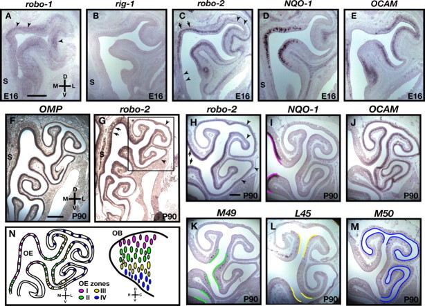
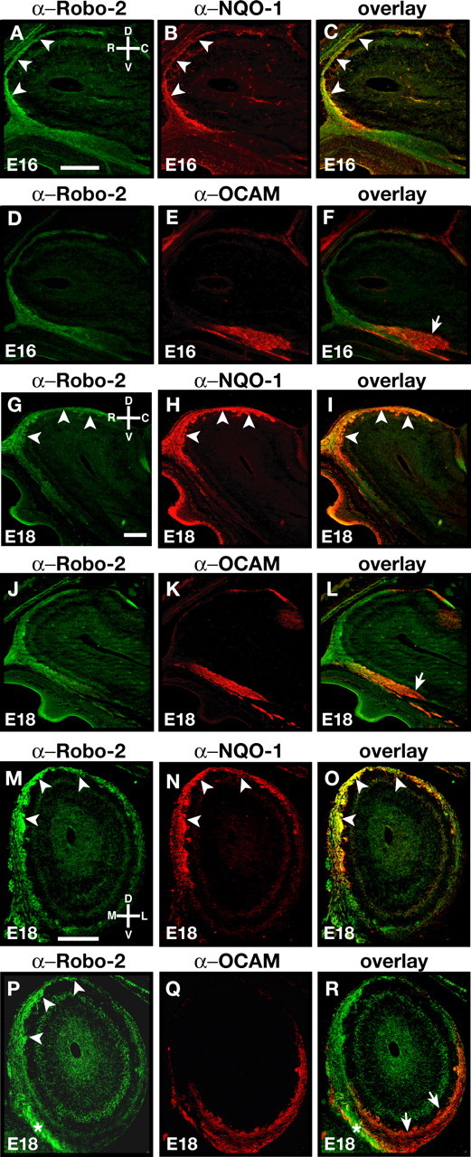
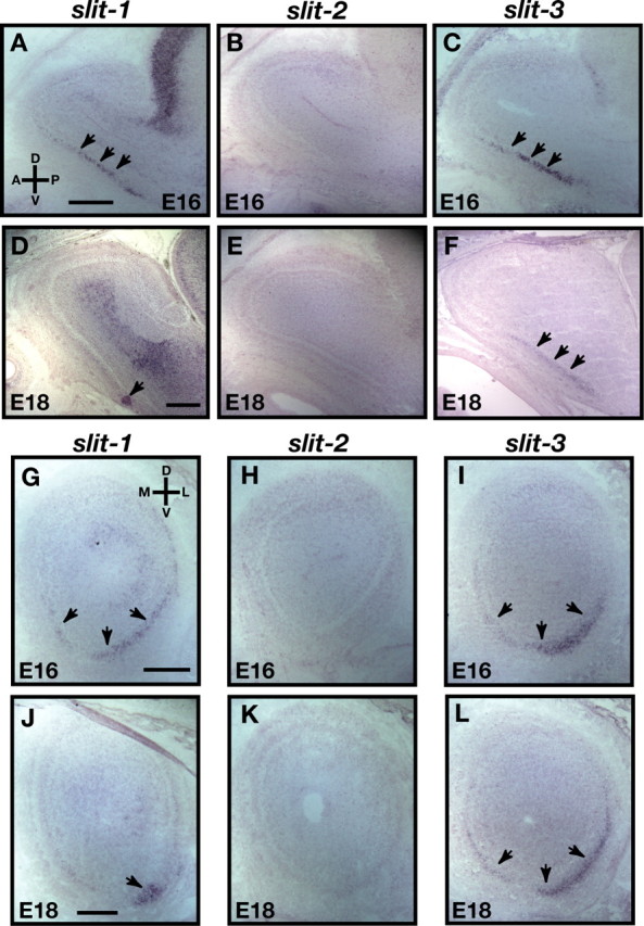
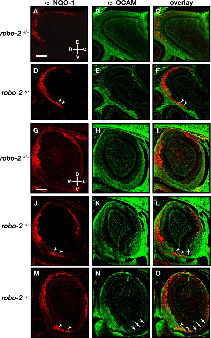
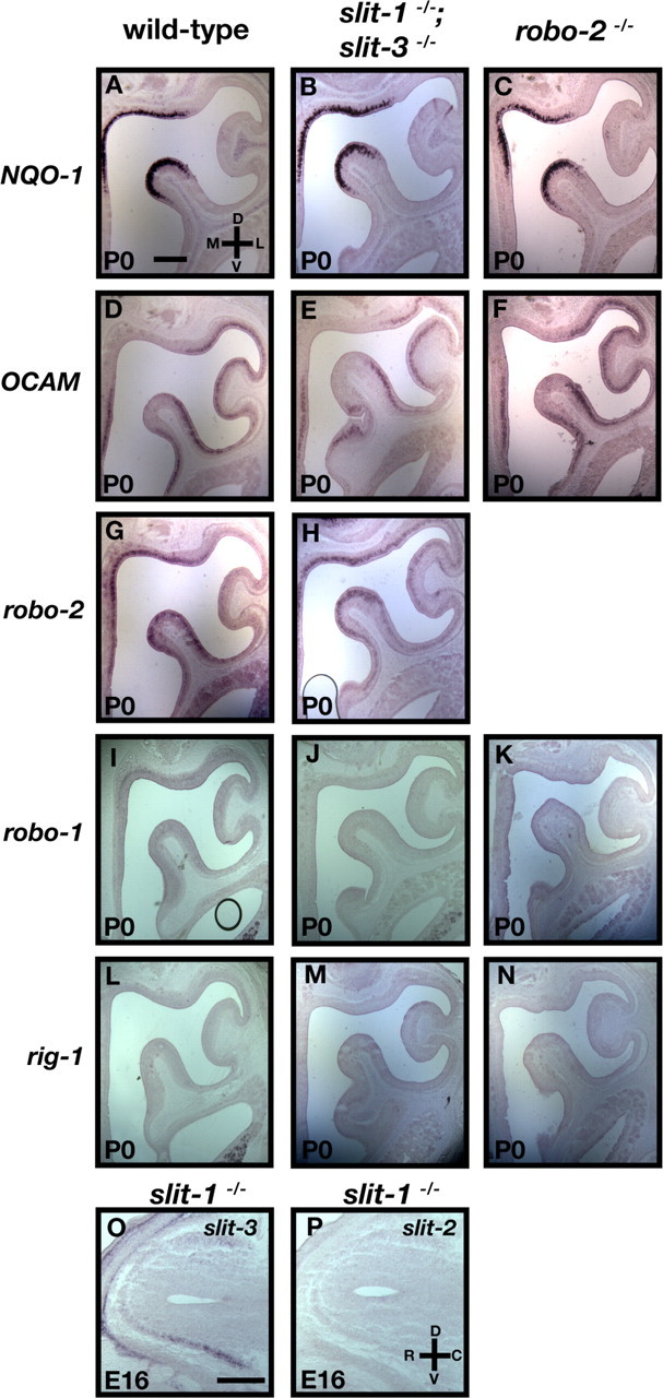
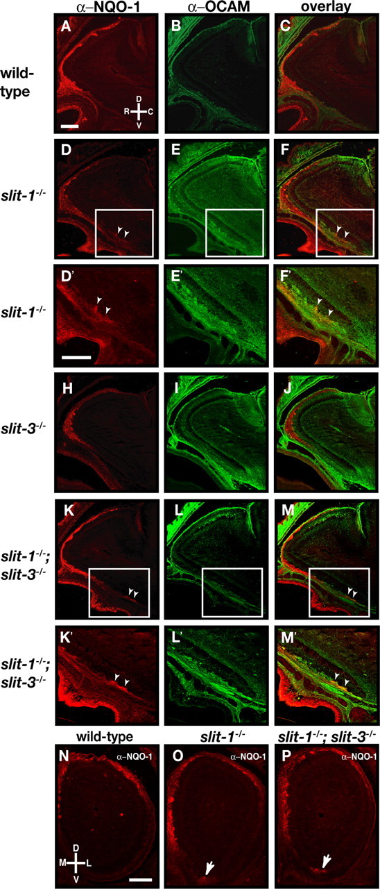
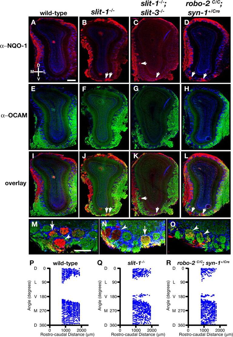
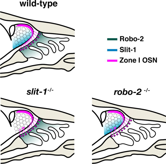
The formation of precise stereotypic connections in sensory systems is critical for the ability to detect and process signals from the environment. In the olfactory system, olfactory sensory neurons (OSNs) project axons to spatially defined glomeruli within the olfactory bulb (OB). A spatial relationship exists between the location of OSNs within the olfactory epithelium (OE) and their glomerular targets along the dorsoventral axis in the OB. The molecular mechanisms underlying the zonal segregation of OSN axons along the dorsoventral axis of the OB are poorly understood. Using robo-2(-/-) (roundabout) and slit-1(-/-) mice, we examined the role of the Slit family of axon guidance cues in the targeting of OSN axons during development. We show that a subset of OSN axons that normally project to the dorsal region of the OB mistarget and form glomeruli in the ventral region in robo-2(-/-) and slit-1(-/-) mice. In addition, we show that the Slit receptor, Robo-2, is expressed in OSNs in a high dorsomedial to low ventrolateral gradient across the OE and that Slit-1 and Slit-3 are expressed in the ventral region of the OB. These results indicate that the dorsal-to-ventral segregation of OSN axons are not solely defined by the location of OSNs within the OE but also relies on axon guidance cues.


Figures

Figure 1.
Figure 1.
Expression of Robo family members in the olfactory epithelium. In situ hybridization of coronal sections of olfactory epithelium isolated from E16 embryos (A–E) and adult mice (P90) (F–M) with cRNA probes specific for robo-1 (A), rig-1 (B), robo-2 (C, G, H), NQO-1 (D, I), OCAM (E, J), OMP (F), M49 (K), L45 (L), and M50 (M). A–G, Expression of robo-1 is restricted to the basal lamina of the olfactory epithelium (A, arrowheads), whereas rig-1 is not expressed in the olfactory epithelium (B). Interestingly, in contrast to OMP, which is equally expressed throughout the olfactory epithelium, robo-2 is expressed in a gradient with high levels of expression in the dorsomedial regions of the olfactory epithelium (arrows) and low levels in the ventrolateral regions (arrowheads) of E16 (C) and adult olfactory epithelium (G, H). H–M, robo-2 expression is confined to zones I–III of the olfactory epithelium. A higher magnification of a region of the olfactory epithelium (boxed in G) in which all four zones of the olfactory epithelium are represented is shown in H–M. The expression pattern of robo-2 was compared with the expression pattern of specific olfactory epithelium zonal markers that include NQO-1 (zone I) (I), OCAM (zones II–IV) (J), M49 (zone II) (K), L45 (zone III) (L), and M50 (zone IV) (M). robo-2 is expressed in a high-to-low gradient in olfactory sensory neurons located in zones I (arrows) to IV (arrowheads), respectively. Regions of the olfactory epithelium expressing the different zonal markers are traced with a colored line on the apical surface of the olfactory epithelium to represent the four zones (zone I, magenta; zone II, green; zone III, yellow; zone IV, blue). S, Septum. Scale bars: A–E, H–M, 250 μm; F, G, 500 μm. N, Diagram representing the spatial relationship between the location of olfactory sensory neurons within the OE and their target glomeruli within the OB. Olfactory sensory neurons located in the dorsomedial regions of the olfactory epithelium (magenta) project axons to glomeruli in the dorsal region of the olfactory bulb, whereas olfactory sensory neurons in the ventrolateral region of the olfactory epithelium (blue and not shown) project axons to the ventral aspect of the olfactory bulb. D, Dorsal; V, ventral; L, lateral; M, medial; R, rostral; C, caudal.
Figure 2.
Figure 2.
Targeting of Robo-2-expressing olfactory sensory neuron axons in the olfactory bulb. A–L, Parasagittal sections of olfactory bulbs from E16 (A–F) and E18 (G–L) embryos stained with anti-Robo-2 (A, C, D, F, G, I, J, L), anti-NQO-1 (B, C, H, I), and anti-OCAM (E, F, K, L). At E16 and E18, olfactory sensory neuron axons expressing high levels of Robo-2 are observed in the dorsal and rostral regions of the OB (A, D, G, J). Robo-2 is also expressed at lower levels on a subset of axons targeting the ventral region of the olfactory bulb. NQO-1-expressing axons, originating from zone I of the olfactory epithelium, that target to the dorsal region of the OB express high levels of Robo-2 (B, C, H, I) (arrowheads). A subset of OCAM-expressing axons that originate from zones II–IV of the olfactory epithelium and target to the ventral region of the olfactory bulb do not express Robo-2 (arrow) (E, F, K, L). M–R, Coronal sections of olfactory bulbs from E18 embryos stained with anti-Robo-2 (M, O, P, R), anti-NQO-1 (N, O), and anti-OCAM (Q, R). At E18, olfactory sensory neuron axons expressing high levels of Robo-2 are observed in the dorsomedial regions of the OB (arrowheads), whereas lower levels of Robo-2 expression are observed on olfactory sensory neuron axons targeting to the ventrolateral region of the OB. As observed in sagittal sections (G–L), NQO-1-positive axons express high levels of Robo-2 (arrowheads) (M–O), whereas a subset of OCAM-positive axons do not express Robo-2 (arrows) (P–R). Robo-2-positive axons restricted to the nerve layer are marked with asterisks in P and R. D, Dorsal; V, ventral; R, rostral; C, caudal; M, medial; L, lateral. Scale bars, 250 μm.
Figure 3.
Figure 3.
Expression of slits in the olfactory bulb. A–L, In situ hybridization of parasagittal (A–F) and coronal (G–L) sections of olfactory bulb at E16 (A–C, G–I) and E18 (D–F, J–L) with cRNA probes specific for slit-1 (A, D, G, J), slit-2 (B, E, H, K), and slit-3 (C, F, I, L). At E16, whereas slit-2 is not expressed in the olfactory bulb (B, H), slit-1 (A, G) and slit-3 (C, I) are expressed in the ventral region of the olfactory bulb (arrows). These patterns of expression are maintained at E18 for slit-2 (E, K) and slit-3 (F, L), whereas slit-1 expression is restricted to the most ventral region of the OB at E18 (D, J) (arrows). D, Dorsal; V, ventral; L, lateral; M, medial; R, rostral; C, caudal. Scale bars, 250 μm.
Figure 4.
Figure 4.
Olfactory sensory neuron projections are disorganized in robo-2−/− mice. Parasagittal (A–F) and coronal (G–O) sections of olfactory bulbs from P0 robo-2+/+ (A–C, G–I) and robo-2−/− (D–F, J–O) mice were stained with anti-NQO-1 (A, C, D, F, G, I, J, L, M, O) and anti-OCAM (B, C, E, F, H, I, K, L, N, O). In robo-2−/− mice, NQO-1-expressing axons are restricted to the rostral and dorsal region of the olfactory bulbs (A, G), whereas OCAM-expressing axons target the ventral region of the olfactory bulbs (B, H). A subset of NQO-1-expressing axons is mistargeted to the ventral region of the olfactory bulbs in robo-2−/− mice (arrowheads) (D, F, J, L, M, O). In addition, some regions of the ventral olfactory bulb that are innervated by OCAM-expressing axons in robo-2+/+ mice lack innervation in robo-2−/− mice (arrows). n = 9 robo-2+/+, 3 robo-2+/−, and 11 robo-2−/−. D, Dorsal; V, ventral; L, lateral; M, medial; R, rostral; C, caudal. Scale bars, 250 μm.
Figure 5.
Figure 5.
Expression of NQO-1 and OCAM are unaltered in robo-2−/− and slit-1−/−;slit-3−/− mice. A–N, In situ hybridization of coronal sections of olfactory epithelia from P0 wild-type (A, D, G, I, L), slit-1−/−;slit-3−/− (B, E, H, J, M), and robo-2−/− (C, F, K, N) mice with cRNA probes specific for NQO-1 (A–C), OCAM (D–F), robo-2 (G, H), robo-1 (I–K), and rig-1 (L–N). robo-2 is expressed in a gradient in the OE with high levels of expression in the dorsomedial region of the OE and low levels in the ventrolateral region in wild-type (G) and slit-1−/−;slit-3−/− (H) mice. NQO-1 is expressed in the dorsomedial region, whereas OCAM is expressed in the ventrolateral region of the olfactory epithelium in wild-type (A, D), slit-1−/−;slit-3−/− (B, E), and robo-2−/− (C, F) mice. Both robo-1 and rig-1 are not expressed in the olfactory epithelium in wild-type (I, L), slit-1−/−;slit-3−/− (J, M), and robo-2−/− (K, N) mice. n = 5 robo-2−/− and n = 5 slit-1−/−;slit-3−/−. O, P, In situ hybridization of parasagittal sections of olfactory bulbs from E16 slit-1−/− embryos with cRNA probes specific for slit-3 (O) and slit-2 (P). Whereas slit-3 is expressed in the ventral region of the olfactory bulb, slit-2 is not expressed in the olfactory bulb of slit-1−/− embryos. n = 3 slit-1−/−. D, Dorsal; V, ventral; L, lateral; M, medial; R, rostral; C, caudal. Scale bars, 250 μm.
Figure 6.
Figure 6.
Loss of zonal targeting of NQO-1-expressing olfactory sensory neuron axons within the olfactory bulb in slit-1−/− mice. A–M, Parasagittal sections of olfactory bulbs from P0 wild-type (A–C), slit-1−/− (D–F), slit-3−/− (H–J), and slit-1−/−;slit-3−/− (K–M) mice stained with anti-NQO-1 (A, C, D, F, H, J, K, M) and anti-OCAM (B, C, E, F, I, J, L, M). In wild-type animals, NQO-1-expressing axons are restricted to the rostrodorsal region of the OB (A, C) and OCAM-expressing axons target to the ventral region of the olfactory bulb (B, C). Whereas NQO-1-expressing axons are properly targeted to the dorsal region of the olfactory bulb in slit-3−/− mice (H, J), a subset of NQO-1-expressing axons mistarget to the most ventral region of the olfactory bulb in slit-1−/− mice (D, F) (arrowheads). In slit-1−/−; slit-3−/− mice, NQO-1-expressing axons are also observed in the ventral region of the olfactory bulb (K, M) (arrowheads). High-powered magnifications of ectopically projecting NQO-1-expressing axons (insets in D, F, K, M) are shown in D′, F′, K′, and M′. n = 8 wild type, n = 5 slit-1−/−, n = 8 slit-3−/−, and n = 7 slit-1−/−; slit-3−/−. N–P, Coronal sections at similar rostrocaudal levels of olfactory bulbs isolated from P0 (N–P) wild-type (N), slit-1−/− (O), and slit-1−/−; slit-3−/− (P) mice stained with anti-NQO-1 (N–P). In wild-type mice, NQO-1-expressing axons innervate the dorsomedial region of the olfactory bulb. In slit-1−/− and slit-1−/−; slit-3−/− mice, a subset of NQO-1-expressing axons are mistargeted to the ventral region of the olfactory bulb (arrows). n = 8 wild type, n = 8 slit-1−/−, and n = 5 slit-1−/−; slit-3−/−. D, Dorsal; V, ventral; L, lateral; M, medial; R, rostral; C, caudal. Scale bars, 250 μm.
Figure 7.
Figure 7.
Mistargeted axons of NQO-1-expressing olfactory sensory neurons innervate glomeruli in the ventral region of the olfactory bulb in slit and robo-2 mutant mice. Coronal sections at similar rostrocaudal levels of olfactory bulbs isolated from 8- to 12-week-old wild-type (A, E, I), slit-1−/− (B, F, J, M–O), slit-1−/−;slit-3−/− (C, G, K), and robo-2c/c;syn-1Cre/+ (D, H, L) mice stained with anti-NQO-1 (A–D, I–L, M–O), anti-OCAM (E–H, I–L, M, N), anti-SV2 (O), and Hoechst (A–O). In wild-type mice, NQO-1-expressing axons innervate glomeruli restricted to the dorsomedial region of the olfactory bulb (A, I), and OCAM-expressing axons innervate glomeruli in the ventrolateral region of the olfactory bulb (E, I). In slit-1−/− (B, J), slit-1−/−;slit-3−/− (C, K), and robo-2c/c;syn-1Cre/+ (D, L) mice, a subset of NQO-1-expressing axons are mistargeted to glomeruli in the ventral region of the olfactory bulb (arrows). NQO-1-expressing axons that target ectopically in the ventral region of the OB in slit-1−/− mice form both homogenous (arrowheads) and heterogenous (arrows) glomeruli (M, N) and are positive for the presynaptic marker SV2, suggesting that they form synapses (arrowheads) (O). n = 10 wild type, n = 14 slit-1−/−, n = 9 slit-1−/−;slit-3−/−, and n = 4 robo-2c/c;syn-1Cre/+. Scale bars: A–L, 250 μm; M–O, 140 μm. P–R, Scatter plots showing the mapping of the positions of NQO-1-positive glomeruli in the olfactory bulb of an adult wild-type (P), slit-1−/− (Q), and robo-2c/c;syn-1Cre/+ (R) mouse. The relative positions of glomeruli containing NQO-1-positive axons were assessed in olfactory bulb sections isolated over a rostrocaudal distance of 1000 μm starting at 800 μm from the tip of the olfactory bulb. Although NQO-1-positive glomeruli are absent in the ventral region of the olfactory bulb (180° angle) from a wild-type mouse, NQO-1-positive glomeruli are consistently observed in the most ventral region of the OB in slit-1−/− and robo-2c/c;syn-1Cre/+ mice. Shown are representative plots from a single olfactory bulb for each genotype (n = 4 olfactory bulbs from 2 mice of each genotype). D, Dorsal; V, ventral; L, lateral; M, medial.
Figure 8.
Figure 8.
Slit-1 and Robo-2 are required for the segregation of zone I OSN axons to the dorsal region of the OB. Representation of zone I OSN projections in wild-type, slit-1−/−, and robo-2−/− mice. NQO-1-expressing zone I OSNs (magenta) located in the dorsal region of the OE project their axons to the dorsal aspect of the OB. The high dorsal to low ventral graded expression of Robo-2 (green) in OSN of the OE promotes the segregation of NQO-1-expressing axons to the dorsal region of the OB. Zone I OSN axons may be repelled by the Robo-2 ligand Slit-1, which is expressed in the ventral region of the OB. Loss of either Robo-2 or Slit-1 expression leads to mistargeting of a subset of zone I OSN axons to the ventral aspect of the OB.

Similar articles

Cited by

References

    1. Alenius M, Bohm S. Identification of a novel neural cell adhesion molecule-related gene with a potential role in selective axonal projection. J Biol Chem. 1997;272:26083–26086. - PubMed
    1. Astic L, Saucier D. Anatomical mapping of the neuroepithelial projection to the olfactory bulb in the rat. Brain Res Bull. 1986;16:445–454. - PubMed
    1. Brose K, Bland KS, Wang KH, Arnott D, Henzel W, Goodman CS, Tessier-Lavigne M, Kidd T. Slit proteins bind Robo receptors and have an evolutionarily conserved role in repulsive axon guidance. Cell. 1999;96:795–806. - PubMed
    1. Chesler AT, Zou D-J, Le Pichon CE, Peterlin ZA, Matthews GA, Pei X, Miller MC, Firestein S. A G protein/camp signal cascade is required for axonal convergence into olfactory glomeruli. Proc Natl Acad Sci USA. 2007;104:1039–1044. - PMC - PubMed
    1. Cloutier J-F, Giger RJ, Koentges G, Dulac C, Kolodkin AL, Ginty DD. Neuropilin-2 mediates axonal fasciculation, zonal segregation, but not axonal convergence, of primary accessory olfactory neurons. Neuron. 2002;33:877–892. - PubMed

Publication types

MeSH terms

LinkOut - more resources