. 2007 Aug 22;27(34):9115-29.
doi: 10.1523/JNEUROSCI.2361-07.2007.

Immunotherapy targeting pathological tau conformers in a tangle mouse model reduces brain pathology with associated functional improvements

Affiliations

Immunotherapy targeting pathological tau conformers in a tangle mouse model reduces brain pathology with associated functional improvements

Ayodeji A Asuni et al. J Neurosci. .

Abstract

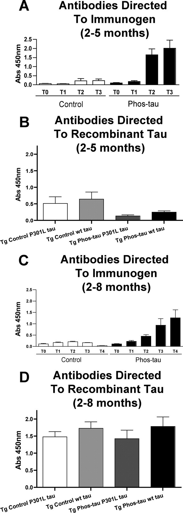
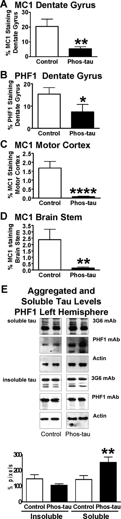
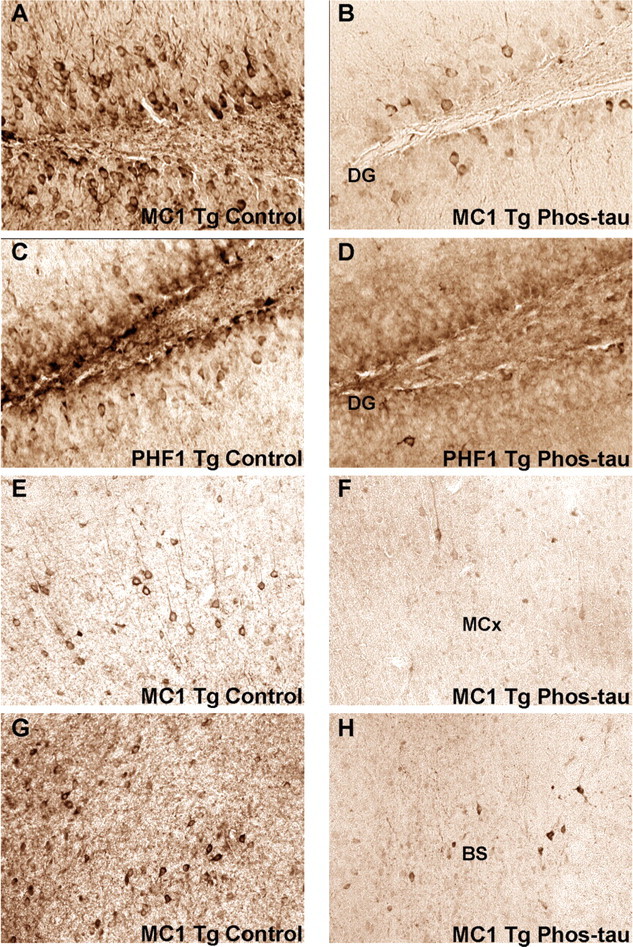
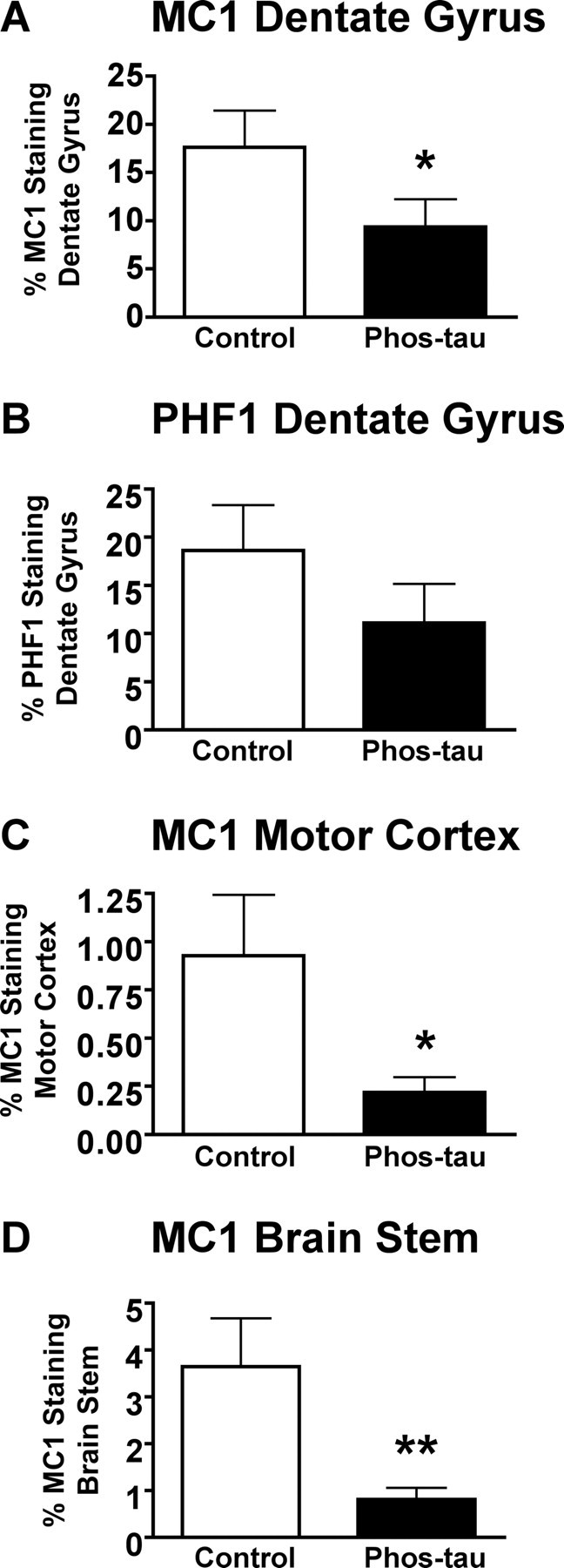
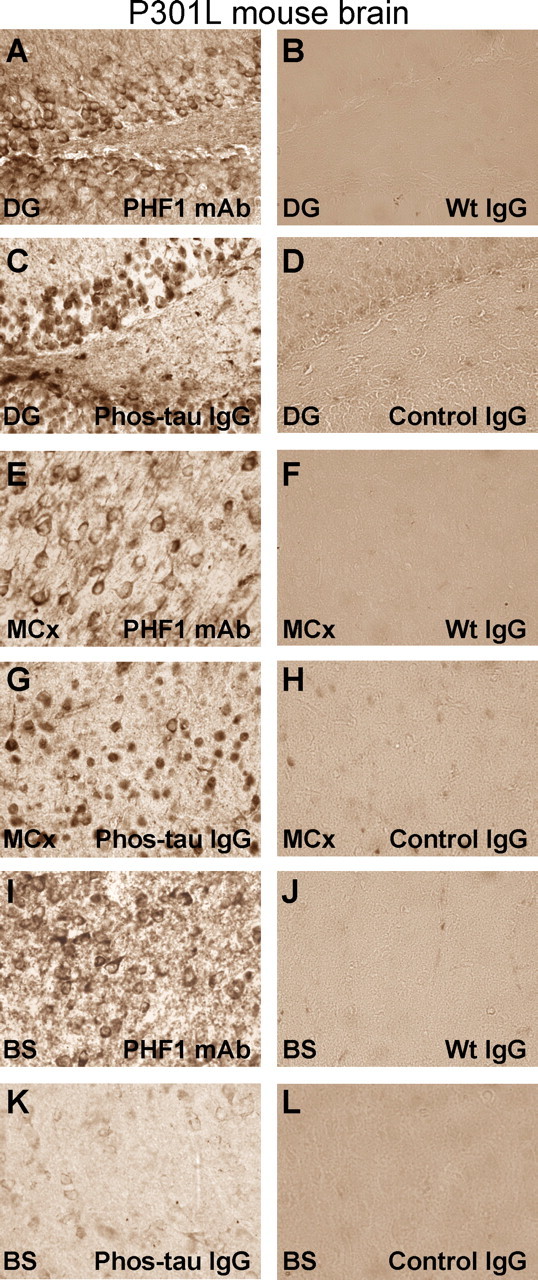
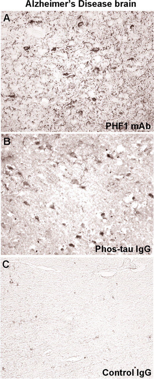
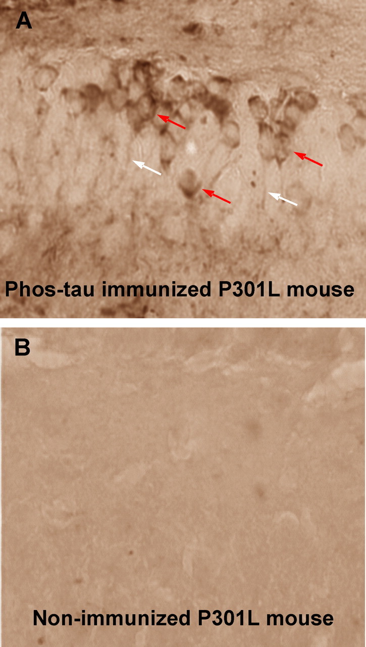
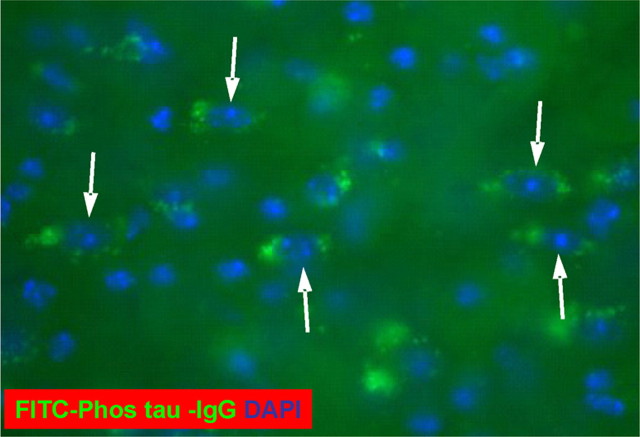
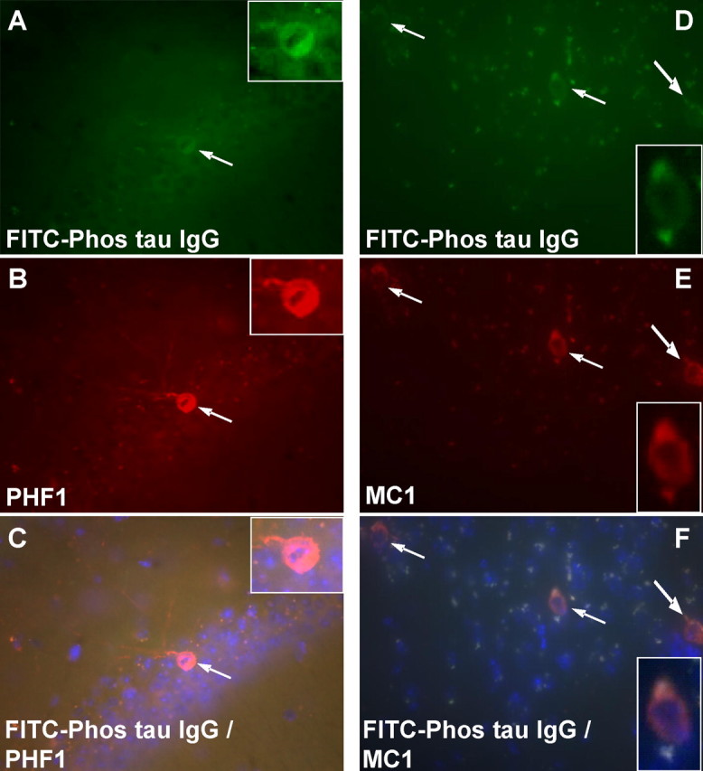
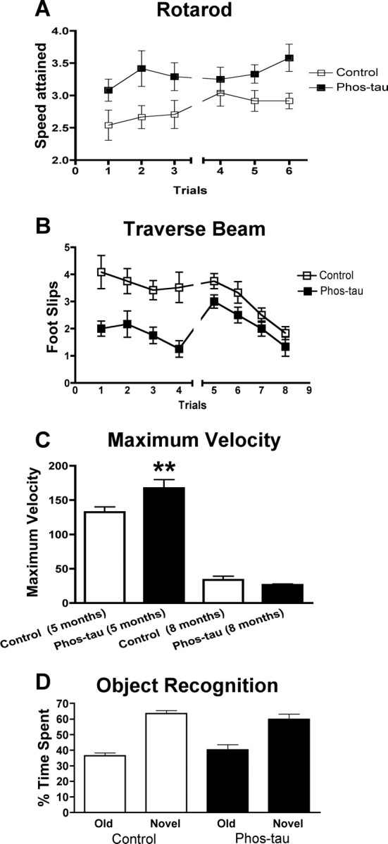
Immunotherapies for various neurodegenerative diseases have recently emerged as a promising approach for clearing pathological protein conformers in these disorders. This type of treatment has not been assessed in models that develop neuronal tau aggregates as observed in frontotemporal dementia and Alzheimer's disease. Here, we present that active immunization with a phosphorylated tau epitope, in P301L tangle model mice, reduces aggregated tau in the brain and slows progression of the tangle-related behavioral phenotype. Females had more tau pathology than males but were also more receptive to the immunotherapy. The tau antibodies generated in these animals recognized pathological tau on brain sections. Performance on behavioral assays that require extensive motor coordination correlated with tau pathology in corresponding brain areas, and antibody levels against the immunogen correlated inversely with tau pathology. Interestingly, age-dependent autoantibodies that recognized recombinant tau protein but not the immunogen were detected in the P301L mice. To confirm that anti-tau antibodies could enter the brain and bind to pathological tau, FITC-tagged antibodies purified from a P301L mouse, with a high antibody titer against the immunogen, were injected into the carotid artery of P301L mice. These antibodies were subsequently detected within the brain and colocalized with PHF1 and MC1 antibodies that recognize pathological tau. Currently, no treatment is available for clearing tau aggregates. Our present findings may lead to a novel therapy targeting one of the major hallmarks of Alzheimer's disease and frontotemporal dementia.


Figures

Figure 1.
Figure 1.
Phospho-tau derivative peptide is highly immunogenic in P301L mice, but autoantibodies against tau are detected. Homozygous Tg P301L mice were immunized from 2 months of age with a phosphorylated tau peptide (Phos-tau, Tau379–408[P-Ser396,404]; n = 24). Control Tg P301L animals received aluminum adjuvant alone (n = 25). Plasma samples from the animals were analyzed by ELISA, and the brains were analyzed biochemically and immunohistochemically at 5 months of age (Phos-tau, n = 14; controls, n = 12) and at 8 months of age (Phos-tau, n = 12; controls, n = 12). A, C, Shown is the generation of IgG antibodies (1:200 plasma dilution) against the immunogen at various time points as determined by Phos-tau peptide ELISA assay [A, 2–5 months: T0, T1, T2, T3 = 0, 7, 11, and 14 weeks; C, 2–8 months: T0, T1, T2, T3, T4 = 0, 7, 11, 14, and 26 weeks]. Control mice had low levels of autoantibodies that recognized the immunogen. B, D, Autoantibodies that recognized both wild-type and P301L human tau were observed in controls and immunized mice within both groups at the end of each study (B, 2–5 months; D, 2–8 months), but the levels did not differ significantly between the groups. Similar reactivity was observed toward wild-type and P301L human tau. Error bars indicate SEM.
Figure 2.
Figure 2.
The vaccine reduces tau aggregates in the brains of P301L tangle mice at 5 months of age. A, Quantitative analysis of MC1 immunoreactivity within the granular layer of the dentate gyrus revealed a 74% reduction (**p < 0.01) in immunized mice compared with control Tg mice that received adjuvant alone. B, Likewise, PHF1 immunoreactivity within the granular layer of the dentate gyrus was reduced by 52% (*p < 0.05) in immunized mice compared with controls. C, Additional confirmation of a therapeutic effect was obtained by analysis of the motor cortex, in which MC1 neuronal staining was reduced by 96% (****p < 0.0001) compared with control Tg mice. D, Likewise, in the brainstem, MC1 neuronal staining was reduced by 93% (**p = 0.01) compared with control Tg mice. E, Densitometric analysis of PHF1 blots revealed a strong trend for reduction in insoluble tau (28% reduction; p = 0.09) and a significant increase in soluble tau (77% increase; **p = 0.01) in the immunized mice compared with control Tg mice, relative to total tau levels. Additional analysis of the ratio of soluble tau to insoluble tau indicated a significant increase in the immunized group on PHF1 blots (89% increase; p = 0.01), suggesting a mobilization of tau from its insoluble form to soluble form in these treated animals. The panel shows representative blots from control and Phos-tau-immunized mice. The PHF1 antibody recognizes phosphorylated serines 396 and 404 located outside the microtubule-binding repeat on the C terminus of PHF tau protein. Antibodies against total tau (3G6) and actin were used as controls. The same amount of protein was loaded in each line. Mean values are presented with SEM.
Figure 3.
Figure 3.
The immunotherapy reduces pathological tau in neurons. Representative examples of the histological regions that were analyzed in MC1- and PHF1-stained brain sections of mice at 5 months in the 2–5 month study group. Neuronal tau aggregates were cleared in the dentate gyrus (DG), the motor cortex (MCx), and the brainstem (BS) in immunized mice compared with control mice. The dentate gyrus develops extensive tau pathology at an early age in the homozygous P301L mice and tau pathology in the motor cortex and brainstem may relate to the motor deficits in this model. A, B, Coronal section through the dentate gyrus of MC1-stained section in control (A) versus immunized (B) Tg mouse (original magnification, 200×). C, D, Coronal section through the dentate gyrus of PHF1-stained section in a control (C) versus immunized (D) Tg mouse (original magnification, 200×). E, F, Coronal section through the motor cortex of MC1-stained section in a control (E) versus immunized (F) Tg mouse (original magnification, 100×). G, H, Coronal section through the brainstem below the aqueduct of Sylvius of MC1-stained section in a control (G) versus immunized (H) Tg mouse (original magnification, 100×). The following magnifications were used for the quantitative analysis: dentate gyrus, 200×; motor cortex, 100×; brainstem, 100×.
Figure 4.
Figure 4.
Immunotherapy from 2 to 8 months reduces brain tau pathology. A, Quantitative analysis of MC1 immunoreactivity within the granular layer of the dentate gyrus revealed a 47% reduction (*p < 0.05) in immunized mice compared with control Tg mice that received adjuvant alone. B, There was a strong trend for a diminished PHF1 immunoreactivity within the granular layer of the dentate gyrus (40% reduction; p < 0.12) in immunized mice compared with controls. C, As at 5 months of age (Fig. 2C), MC1 neuronal staining of the motor cortex revealed a more pronounced therapeutic effect (76%; *p = 0.02) than in the dentate gyrus. D, Likewise, neuronal MC1 staining was substantially reduced in the brainstem of immunized mice compared with controls (78%; **p = 0.005). Mean values are presented with SEM.
Figure 5.
Figure 5.
Purified antibodies from immunized mice stain tau aggregates/tangles in neuronal cell bodies in P301L mice. A–L, Adjacent coronal brain sections through the dentate gyrus (A–D), motor cortex (E–H), and brainstem (I–L; immediately below the Aqueduct of Sylvius) in a P301L transgenic mouse with tau pathology. A, The PHF1 antibody reveals the typical staining of tau aggregates/tangles in neuronal cell bodies as previously reported in this model (Lewis et al., 2000). Most of the staining is associated with the neuronal plasma membrane. B, D, Pooled mouse IgG from wild-type mice [Wt IgG (Sigma)] (B) or antibodies from mice immunized with the adjuvant only (Control IgG) (D) do not decorate neurons in the dentate gyrus. C, Antibodies from mice immunized with the Phos-tau peptide, which contains the PHF1 epitope, lack extensive dendritic staining, but stain primarily neuronal cell bodies within the dentate gyrus, but the pattern is not identical to the PHF1 staining. E, G, I, K, Similar staining pattern as in A and C is obsered in the motor cortex (E, G) and brainstem (I, K) after immunoreactivity with the PHF1 antibody and the polyclonal antibody from an immunized mouse, respectively. However, this particular polyclonal antibody stained neurons in the brainstem less intensely than in the dentate gyrus and motor cortex. F, H, J, L, These images depict adjacent coronal sections to those shown in E, G, I, and K, which were stained with purified antibodies from Tg control mice that received adjuvant alone (Control IgG) or pooled mouse IgG (Wt IgG). Staining with those antibodies resulted in minimal or no staining. No immunostaining was observed in wild-type mice with the antibodies purified from immunized mice (data not shown). These findings indicate that the immunized mice generate antibodies that specifically recognize pathological tau aggregates in the P301L mouse. Staining was performed as detailed in Materials and Methods with PHF1 and purified IgG used at a 1:250 and 10 μg/ml dilutions, respectively. Original magnification, 400×.
Figure 6.
Figure 6.
Purified antibodies from immunized mice stain tau aggregates/tangles in neuronal cell bodies in Alzheimer's disease similar to the PHF1 antibody. A, PHF1 staining of the entorhinal cortex from an Alzheimer's brain reveals the typical staining of cell bodies and dystrophic neurites as previously described for this antibody. B, The polyclonal IgG antibody derived from immunized mouse stains neuronal cell bodies, although this was dissimilar to PHF1 monoclonal antibody and dystrophic neurites are not prominent. C, Antibodies purified from a control mouse that received adjuvant alone do not result in appreciable staining. Overall, the staining pattern with these different antibodies is comparable with that observed in the P301L mouse (Fig. 5).
Figure 7.
Figure 7.
Intracerebral antibodies that label neurons are detected in the immunized P301L mice. A, A coronal brain section, representative of a high-titer P301L mouse, stained with an anti-IgG secondary antibody (1:50; Vectastain Elite kit) through the dentate gyrus of the hippocampus. Note the staining of neuronal cell bodies (red arrows) and processes (white arrows) indicating the presence of IgG. No immunostaining is observed in a representative nonimmunized P301L mouse of a similar age (B) or in a wild-type mouse (data not shown) under these staining conditions. Original magnification, 400×.
Figure 8.
Figure 8.
FITC-tagged IgG antibodies from an immunized mouse label neurons in the brain after intracarotid injection into an 8-month-old P301L mouse. A coronal brain section through the brachium of the inferior colliculus revealing FITC-labeled neurons (arrows). Counterstain with DAPI (blue) shows nuclei of the neurons. Some FITC labeling was observed in control P301L mice of the same age that were injected with tagged IgG antibodies from pooled mouse plasma (Sigma), but neurons were not detected (data not shown). No appreciable FITC fluorescence was observed in wild-type mice of the same age that received intracarotid injection of IgG antibodies from an immunized mouse or control IgG (data not shown).
Figure 9.
Figure 9.
Neurons that label with the injected FITC-tagged antibody from an immunized mouse stain with MC1 and PHF1 antibodies. A–C, A coronal brain section through the pyramidal layer of the hippocampus. Note the FITC-labeled neurons in A that stain with PHF1 antibody that was applied to the section (B; Texas Red-tagged secondary antibody). The section was counterstained with DAPI that stains nuclei in blue and double-labeled neurons are orange (C). Original magnification, 200×. D–F, A coronal brain section through the nucleus of the brachium inferior colliculus. Note the FITC-labeled neurons in D that stain with MC1 antibody that was applied to the section (E; Texas Red-tagged secondary antibody). The section was counterstained with DAPI, which stains nuclei in blue, and double-labeled neurons are orange (F). Original magnification, 200×.
Figure 10.
Figure 10.
Immunotherapy from 2 to 8 months of age slows the progression of behavioral abnormalities in P301L mice. A shows the performance of Tg P301L-immunized and Tg control mice, trained to remain on a rotarod, and the speed attained during the task. The immunization increased the time the animals were able to stay on the rotarod both at 5 months (trials 1–3, p < 0.02) and 8 months (trials 4–6, p < 0.05). B shows the number of foot slips the animals had during the traverse beam task. The immunization greatly reduced number of foot slips during the performance of the task at 5 months (p < 0.001) and at 8 months (p = 0.05). C, There was no significant difference in the distance traveled, average speed (Vmean), or the resting time at 5 and 8 months (data not shown), but there was an increase in the maximum velocity (Vmax) attained by the phospho-tau-immunized Tg animals (**p = 0.004) at 5 months, compared with Tg controls. Vmax did not differ between the groups at 8 months. D, No difference was observed between the groups in the object recognition task that measures short-term memory. Both the immunized P301L mice and their transgenic controls spent a comparable time exploring the novel object that differed substantially from the time they spent with the old object. This finding indicates that both groups had normal short-term memory at 8 months of age. Error bars indicate SEM.

References

    1. Aihara N, Tanno H, Hall JJ, Pitts LH, Noble LJ. Immunocytochemical localization of immunoglobulins in the rat brain: relationship to the blood-brain barrier. J Comp Neurol. 1994;342:481–496. - PubMed
    1. Andoh T, Kuraishi Y. Direct action of immunoglobulin G on primary sensory neurons through Fc gamma receptor I. FASEB J. 2004;18:182–184. - PubMed
    1. Andorfer C, Acker CM, Kress Y, Hof PR, Duff K, Davies P. Cell-cycle reentry and cell death in transgenic mice expressing nonmutant human tau isoforms. J Neurosci. 2005;25:5446–5454. - PMC - PubMed
    1. Asuni AA, Knudsen E, Frangione B, Wisniewski T, Sigurdsson EM. Antibody mediated modulation of Aβ induced neurotoxicity in cell culture. Neurobiol Aging. 2004;25:S581–S582.
    1. Asuni AA, Boutajangout A, Scholtzova H, Knudsen E, Li YS, Quartermain D, Frangione B, Wisniewski T, Sigurdsson EM. Vaccination of Alzheimer's model mice with Aβ derivative in alum adjuvant reduces Aβ burden without microhemorrhages. Eur J Neurosci. 2006;24:2530–2542. - PMC - PubMed

Publication types

MeSH terms